8-year-old, female, West Highland White Terrier. A couple of weeks ago, she started with dyschezia / tenesmus along with diarrhea, signs of pollakiuria and weight loss. Abdominal mass identified on Xray and ultrasound.
An abdominal and thoracic CT scan was performed
Description
Abdomen
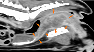
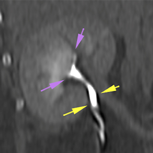
There is a marked and diffuse thickening of the cranial urethral wall, continuing into a mass-like lesion at intrapelvic level (orange arrows). This lesion has irregular and well-defined margins, with a heterogeneous soft tissue attenuation and a marked and moderately heterogeneous contrast enhancement.
At intrapelvic level, the mass causes a marked dorsal displacement of the rectum.
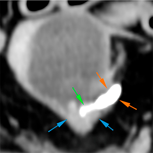
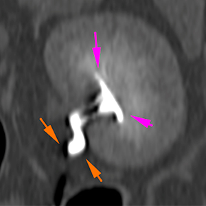
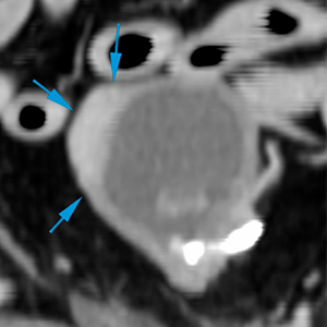
This thickening extends cranially until the trigone of the bladder (blue arrows), where the left ureterovesical junction appears normal (green arrow), although the most distal portion of the left ureter is slightly distended, extending cranially with the same diameter, with a normal path (orange arrows). The left renal pelvis is slightly distended (pink arrows). The right ureterovesical junction, ureter (yellow arrows) and renal pelvis (purple arrow) are unremarkable.
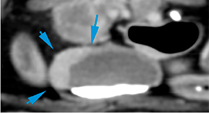
On the right side of the cranial aspect of the bladder, there is an eccentric wall thickening, showing a marked and homogeneous contrast enhancement (blue arrows).
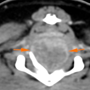
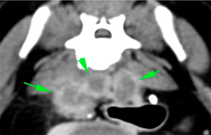
Severe enlargement of the medial and internal iliac lymph nodes, presenting a markedly irregular shape and a heterogeneous contrast enhancement (green arrows).
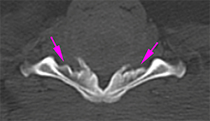
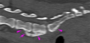
There is a marked new bone formation, with smooth and slightly irregular margins, extending along the pelvic floor (affecting the ilium, pubis, and ischium) as well as along the ventral aspect of the sacrum, and vertebral bodies of L6-L7 (pink arrows).
Thorax
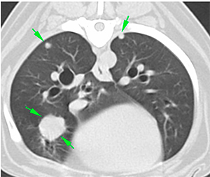
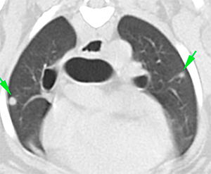
There are multiple, variable in size, soft tissue attenuating nodules, with a mild and homogeneous post-contrast enhancement, distributed throughout the lung fields (green arrows).
Conclusions
Urethral mass of intra-luminal location, extending along the intrapelvic urethra, up to the region of the urinary bladder trigone, consistent with neoplastic disease (transitional cell carcinoma, most likely). The eccentric thickening of the urinary bladder’s wall, at its cranial and right lateral aspect, is consistent with neoplastic infiltration, most likely.
This lesion causes narrowing of the left ureterovesical junction, provoking a mild distention of the ureter and renal pelvis.
The rest of the lesions: marked lumbar lymphadenopathy, periosteal reaction in the vertebral bodies of L6, L7, sacrum, and pelvic bones, as well as the multiple pulmonary nodules, are consistent with metastasis, most likely.
(Mon. to Fri. 9 a.m. to 6 p.m. gmt+1) Welcome, How can we help you?
We use cookies to ensure that we give you the best experience on our website. If you continue to use this site we will assume that you are happy with it.Ok


No comment yet, add your voice below!