Retrobulbar abscess and associated optic neuritis and subdural empyema in a dog
5-year-old female Yorkshire terrier, with conjunctivitis and central vestibular syndrome.
An MRI of the head was performed.
Description
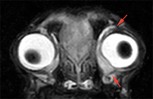
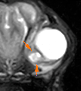
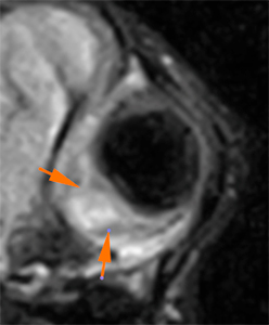
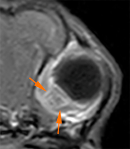
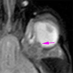
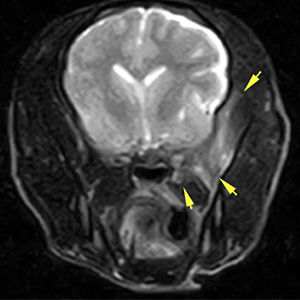
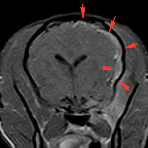
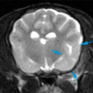
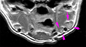
There is a marked thickening of the periorbital fat surrounding the left ocular globe (red arrows). Caudomedial to the left ocular globe, there is a small round lesion, which slightly protrudes towards the ocular globe, causing an indentation in its caudoventral and medial aspect (orange arrows). This lesion is hyperintense on T2W and T2*, with slight signal suppression on FLAIR and hypo/isointense on T1W, with mild peripheral post-contrast enhancement.
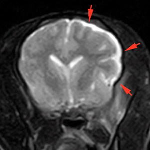
T2W
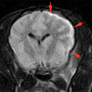
T2W FLAIR T1W+Gd
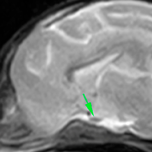
The left optic nerve is slightly thickened, protruding at the level of the optic disc (pink arrow). The optic chiasm appears normal (green arrow).
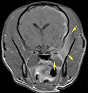
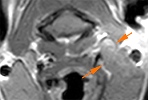
Adjacent to the orbital region, there is a diffuse and marked hyperintensity on T2W, with ill-defined margins, involving the left temporal and medial pterygoid muscles, with marked diffuse post-contrast enhancement (yellow arrows).
T2W T1W+Gd
At intracranial level, adjacent and related to the changes visible at the left orbit, there is a marked thickening of the pachy- and leptomeninges that extends caudally along the entire left cerebral hemisphere, up to the region of the occipital lobe. In addition, there is a crescent-shaped lesion extending along the left cerebral hemisphere hyperintense on T2W and FLAIR and hypointense on T1W, that, after contrast administration, remains hypointense within the meninges.
T1W+Gd T2W FLAIR
In the region of the left temporal lobe, there is a subtle diffuse hyperintensity on T2W affecting the cerebral cortex (blue arrows).
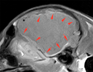
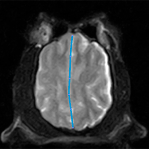
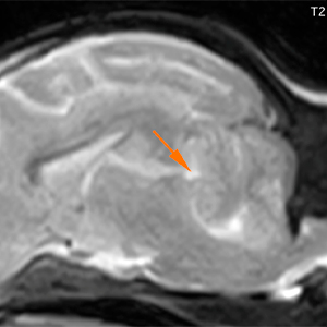
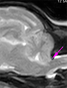
There is a moderate mass effect, with midline shift towards the right (blue line) and a mild transtentorial herniation, causing a moderate protrusion in the rostral aspect of the cerebellum (orange arrow). The caudal portion of the cerebellum slightly protrudes caudally through the foramen magnum (pink arrow).
The ipsilateral mandibular and medial retropharyngeal lymph nodes (pink and orange arrows respectively) are slightly prominent.
Diagnosis
Findings at the level of the left orbit, most likely consistent with left retrobulbar abscess with associated periorbital cellulitis, myositis of the adjacent temporalis and medial pterygoid muscles and left optic neuritis.
Intracranial lesion at the left cerebral hemisphere consistent with meningeal thickening and marked enhancement, as well as associated subdural empyema, most likely, consistent with an infectious process secondary to intracranial extension of the left orbital process. Other ddx, such as meningoencephalitis of unknown origin (GME ocular form) or a neoplastic process are less likely.
Ipsilateral mandibular and medial retropharyngeal lymphadenopathy most likely reactive.
Comments
The findings are consistent with an infectious process affecting the left orbit, with intracranial extension (infectious meningoencephalitis/subdural empyema) most likely, based on the imaging findings, history and blood test abnormalities of the patient.
(Mon. to Fri. 9 a.m. to 6 p.m. gmt+1) Welcome, How can we help you?
We use cookies to ensure that we give you the best experience on our website. If you continue to use this site we will assume that you are happy with it.Ok


2 Comments
Recently we have seen a similar case but we got CT images only.
I can share the case if you think it is interesting…
Cool case! 👍🏽
Hello Maria
Thank you for your comment. It would be great to see those images. You can send them to us at social@protonvet.com