A 10-year-old, female, domestic short hair cat was presented with incoordination. An MRI scan of the head was performed.
Description
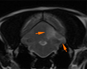
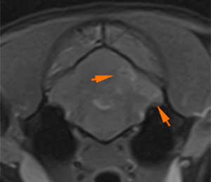
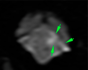
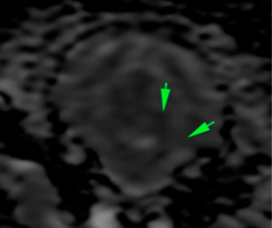
There is an intra-axial lesion affecting mainly the gray matter of the rostral aspect of the left cerebellar hemisphere (orange arrows). The lesion is hyperintense on T2W and FLAIR compared to the grey matter, showing slightly ill-defined margins, hypo/isointense on T1W, with a marked and slightly irregular post-contrast enhancement. On the T2* sequencies, there are no signal voids. The lesion appears hyperintense on DWI and with low values on ADCmap, indicating restricted diffusion (green arrows).
T2W
FLAIR
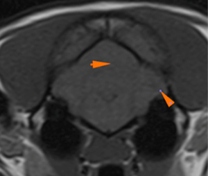
T1W
T1WFS+Gd
T1WFS+Gd
DWI
ADCmap
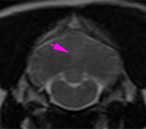
At the level of the caudal aspect of the cerebellum, there is a small intra-axial lesion, showing the same characteristics as the previously described lesion, hyperintense on T2W and FLAIR, isointense on T1W and with moderate contrast enhancement (pink arrows). This lesion cannot be assessed on the diffusion sequences due to its small size.
T2W
FLAIR
T1WFS+Gd
T1WFS+Gd
Diagnosis
The lesion in the rostral aspect of the left cerebellar hemisphere is, most likely, consistent with an ischemic stroke (territorial vascular infarct affecting the left rostral cerebellar artery), based on its characteristics.
Findings in the caudal aspect of the cerebellum are consistent with another ischemic stroke (lacunar infarct), related to the caudal cerebellar artery.
Other differentials, such as a neoplastic process (intravascular lymphoma), could also be possible considering the marked enhancement of the lesions, although less likely.
Comments
The findings in the cerebellum are consistent with ischemic strokes, most likely, considering the characteristics of the lesions and their location. However, it should be considered that the enhancement of these lesions is rare, although it may be possible in both early (24-48h) and later stages (1-8 weeks). A neoplastic process (e.g., intravascular lymphoma) is less likely although it cannot be completely ruled out based on the MRI findings.
CSF analysis is recommended for further investigations. Consider follow-up MRI in several weeks to assess the progression of the lesions. Consider also exclusion of predisposing pathologies for ischemic infarcts (e.g. renal disease, hypertension, etc.).
(Mon. to Fri. 9 a.m. to 6 p.m. gmt+1) Welcome, How can we help you?
We use cookies to ensure that we give you the best experience on our website. If you continue to use this site we will assume that you are happy with it.Ok


No comment yet, add your voice below!