Sacrocaudal intervertebral disc protrusion in a cat
6 years-old, male domestic short hair. Previous history of teeth cleaning one week ago. Now he shows flaccid tail and pain in the lower back and tail area. A spinal CT scan was performed..
Description
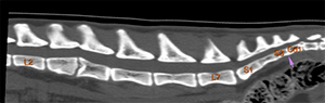
The first coccygeal vertebra (Cd1) is fused with S3 (purple arrows).
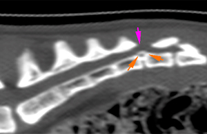
The intervertebral (IV) disc between the first (Cd1) and second (Cd2) coccygeal vertebrae is partially mineralised and moderately protrudes dorsally (orange arrows), causing a narrowing of the vertebral canal (pink arrows).
The IV disc between the fourth (Cd4) and fifth (Cd5) coccygeal vertebrae shows similar changes, being partially mineralized and protruding severely into the vertebral canal. The disc causes complete stenosis of the vertebral canal(orange arrows). The IV discs between the Cd2-Cd3, and Cd3-Cd4 vertebrae, are also partially mineralised, although maintaining their normal position, without causing stenosis of the vertebral canal (blue arrows).
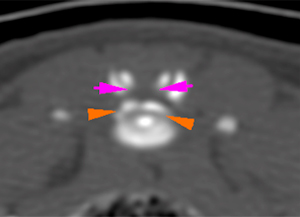
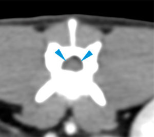
The vertebral bodies of L3 and L4 have a slightly abnormal morphology. The caudal aspect of the vertebral body of L3 and the cranial aspect of the vertebral body of L4 are widened, with very mild irregular endplates and with smooth and well-defined borders (orange arrows). The intervertebral space, at this level, has a slightly irregular/triangular morphology (green arrow). These abnormalities cause minimal stenosis of the vertebral canal, with a very mild kyphosis in the sagittal plane (pink arrowhead). In the vertebral canal, there is a mild dorsal displacement and flattening of the spinal cord, without evident compressive myelopathy (blue arrowheads). There are no alterations in the paraspinal soft tissues.
Diagnosis
1.Extradural lesions in the IV spaces Cd1-Cd2 and Cd4-Cd5, consistent with moderate disc protrusion at Cd1-Cd2 and severe disc protrusion/extrusion at Cd4-Cd5 causing moderate and severe compression of the nerve roots at these levels respectively.
2.Findings at the level of L3 and L4, consistent with a congenital vertebral anomaly or an old lesion affecting the region of the IV disc and endplates (old, non-active discospondylitis). These changes cause minimal stenosis of the vertebral canal, without causing obvious compressive myelopathy. Incidental finding.
3.Partial mineralisation of some IV discs consistent with degenerative changes, without causing compressive myelopathy, considered an incidental finding.
Comments
Findings in the IV spaces of Cd1-Cd2 and Cd4-Cd5 are consistent with degenerative disc disease causing moderate and severe nerve root compression respectively. This could justify the clinical symptomatology of the patient. The rest of the lesions visible in the study are considered incidental without clinical significance. It is recommended to correlate the findings with the neurological examination of the patient.
In the veterinary literature, the most frequently reported locations for intervertebral disc disease (IVDD) among clinically affected cats are T13-L1, L4–L5, and L7-S1. Caudal (coccygeal) and sacrocaudal (sacrococcygeal) IVDD have rarely been reported in cats and clinical signs include pain and neurological dysfunction.
(Mon. to Fri. 9 a.m. to 6 p.m. gmt+1) Welcome, How can we help you?
We use cookies to ensure that we give you the best experience on our website. If you continue to use this site we will assume that you are happy with it.Ok


No comment yet, add your voice below!