Neoplastic process with bilateral hip dysplasia in a dog.
10-years-old, Japanese Spitz. Presented with lameness after another dog ran into him. Radiographs revealed dysplastic hips and a treatment was established. No improvements and slight deterioration were seen after treatment. A CT-scan of the thoracolumbar spine was performed.
Description
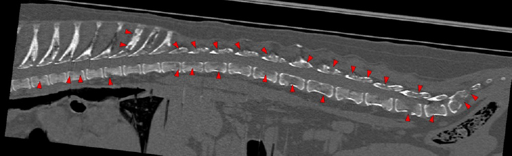
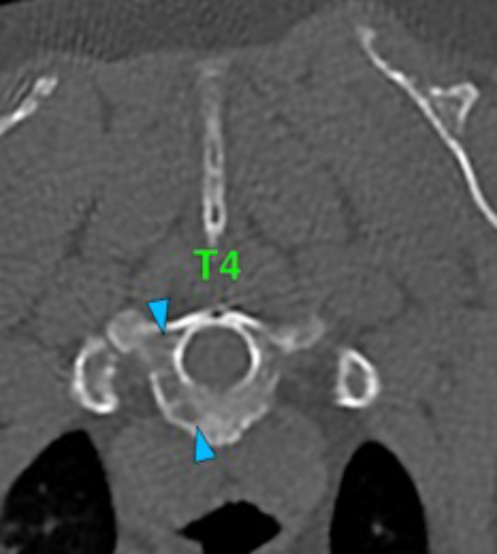
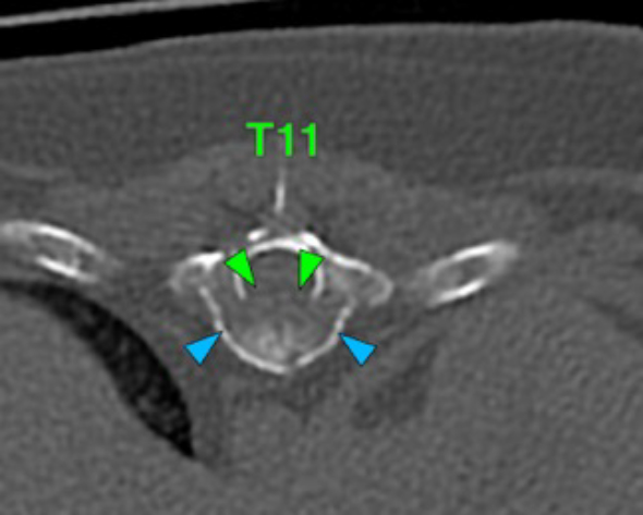
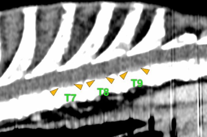
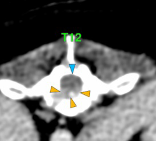
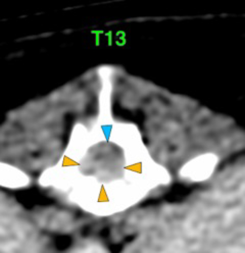
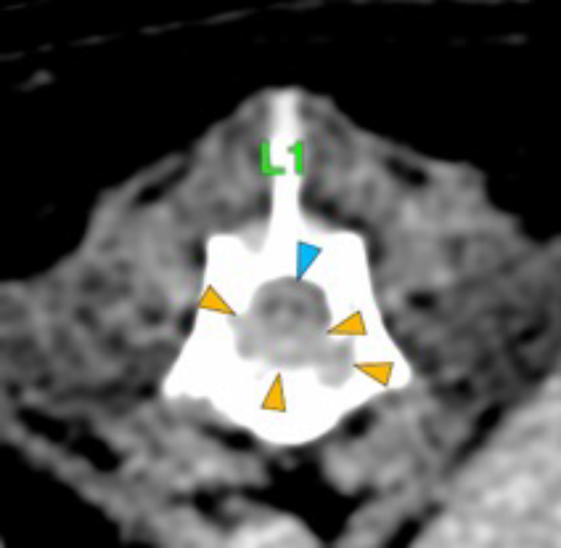
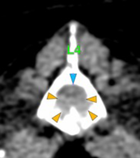
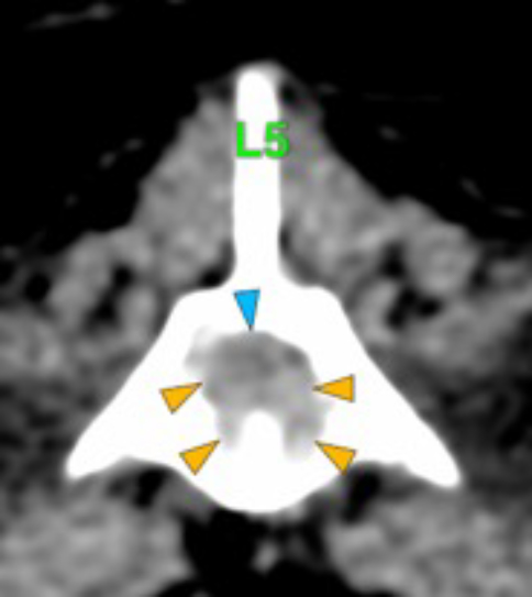
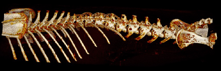
There are multiple osteolytic lesions affecting almost all the vertebral bodies (red arrows). Some of them are areas of permeative or moth-eaten lysis, while others converge into geographic lesions. They affect all the different portions of the vertebra (vertebral body, spinous and transverse processes, etc.) and involve the medullary cavity of the bone (blue arrows), and some of them cause lysis of the cortex (green arrows). Lumbar vertebral bodies more severely affected.
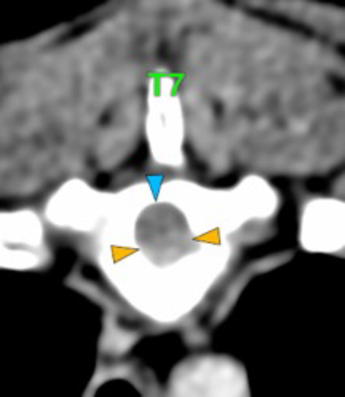
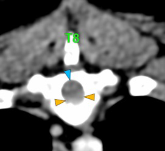
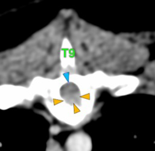
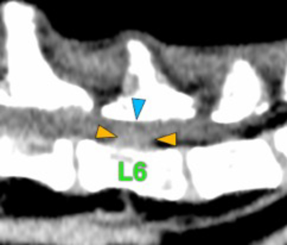
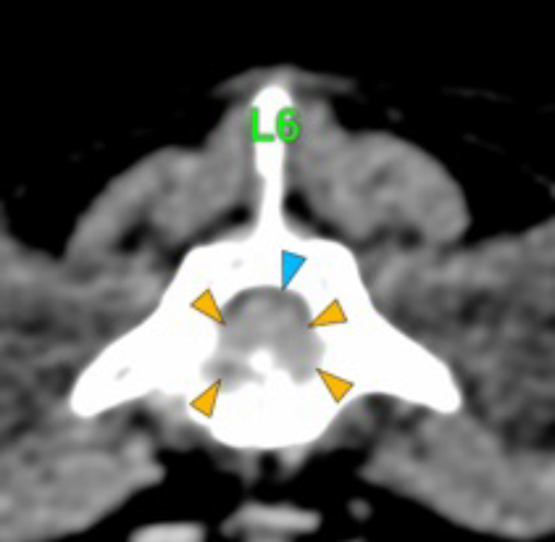
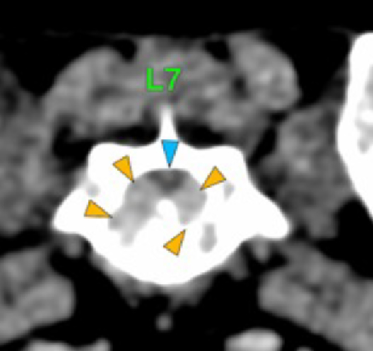
The osteolytic lesions have soft tissue attenuation that shows moderate and slightly heterogeneous post-contrast enhancement. In some of the vertebrae, the lesions cause lysis of the cortex and extend into the vertebral canal (orange arrows) causing variable degree of compression of the spinal cord and cauda equina (blue arrows). At the level of the vertebral bodies of T7, T8, T9 and T12 the lesions extend into the ventral and central aspect of the vertebral canal causing a mild compression of the spinal cord. At the level of T13, L1, L4 and L5 the lesions extend into the ventral and central aspect of the vertebral canal, causing moderate compression of the spinal cord. At the vertebral body of L6 the lesion extends into the ventral and central aspect of the vertebral canal, at the mid-aspect of L6, occupying approx. 90% of the diameter of the vertebral canal causing a severe compression of the cauda equina. At the level of L7, the lesion extends into the ventral and both lateral aspects of the vertebral canal, causing severe compression of the cauda equina.
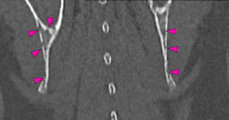
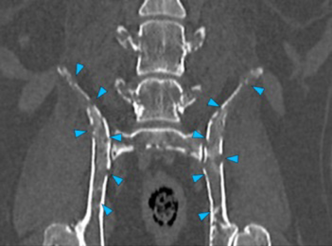
There are multiple osteolytic lesions with the same characteristics affecting both scapula and the pelvic bones included (pink and blue arrows respectively). There are osteolytic lesions with the same characteristics affecting multiple ribs. Associated with these lesions, there are multiple rib fractures.
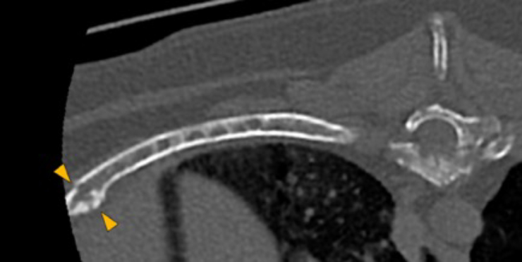
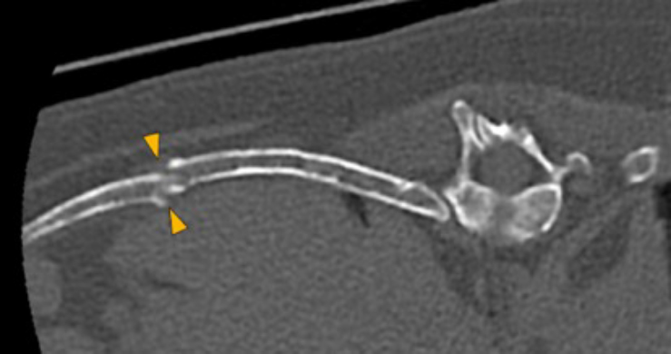
10th and 12th rib
3D image showing the osteolytic lesions described in the skeletal structures included
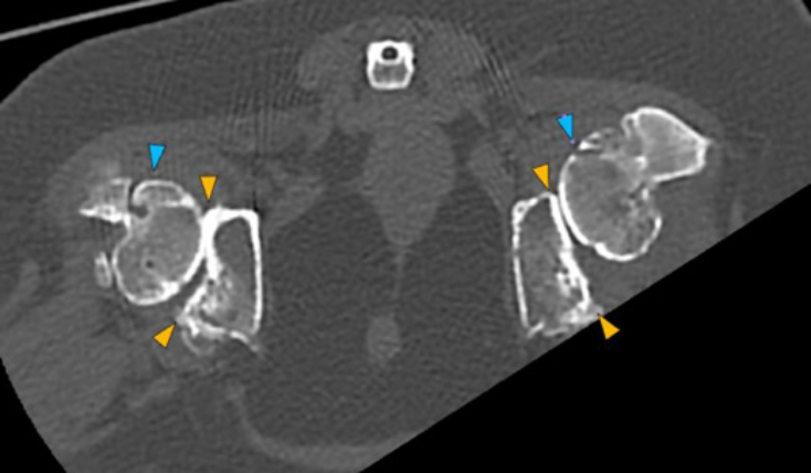
In both coxofemoral joints partially included, there is severe flattening of both acetabula (orange arrows), with marked incongruity of the joint, with dorsal displacement (subluxation) of both femoral heads (blue arrows). In addition, there is severe periarticular new bone formation affecting both acetabula, femoral heads and necks.
Diagnosis
Polyostotic aggressive osseous lesions affecting all the vertebral bodies included, ribs, scapulae and pelvic bones associated with soft tissue lesions showing a moderate and slightly heterogeneous post-contrast enhancement. These changes are most likely consistent with a neoplastic process (multiple myeloma, most likely vs lymphoma or metastasis, less likely). Associated with these lesions, there is:
Soft tissue lesion that extends into the vertebral canal as an extradural lesion in multiple vertebral bodies causing variable degree of compression of the spinal cord and cauda equina (T7-T9 and T12 mild compression of the spinal cord; T13-L1 and L4-L5 moderate compression of the spinal cord; L6 and L7 severe compression of the cauda equina).
Multiple rib fractures.
Changes in both coxofemoral joints consistent with severe degenerative coxofemoral joint disease associated with bilateral hip dysplasia.
(Mon. to Fri. 9 a.m. to 6 p.m. gmt+1) Welcome, How can we help you?
We use cookies to ensure that we give you the best experience on our website. If you continue to use this site we will assume that you are happy with it.Ok

No comment yet, add your voice below!