11-year-old female French Bulldog with edema in the region of the head and neck with increased upper airway sounds. Head, neck, and thoracic CT scan was performed. A thoracolumbar spine and thoracic CT scan was performed.

Description
Thorax
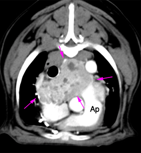
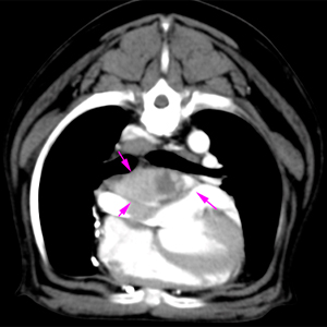
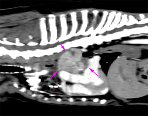
There is a large mass at the base of the heart (up to 6 cm), with irregular and well-defined margins, surrounding the aortic arch. The lesion shows soft tissue attenuation, with marked, heterogeneous contrast enhancement (pink arrows).
This mass causes displacement and compression of the trachea and oesophagus towards the right, of the carina dorsally, and pulmonary arteries and cardiac base ventrally.

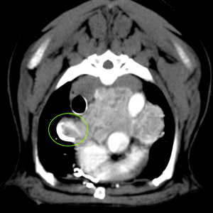
This lesion invades the lumen of the cranial vena cava at the level of the right azygos vein, producing an almost complete obliteration of the lumen and a filling defect (green circle).

There is a small volume of mediastinal effusion (blue arrows).
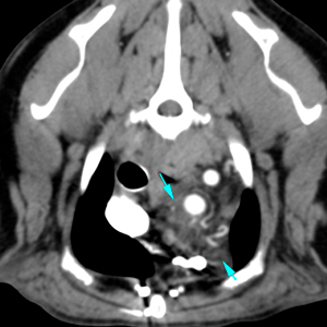
The rest of the cardiovascular structures do not show abnormalities.
No associated regional lymphadenopathy is observed.
The lung parenchyma is unremarkable.
Head
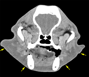
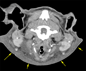
There is a moderate volume of hypoattenuating, non-contrast enhancing fluid (edema) in the facial area, extending ventrally between the deeper muscular planes of the head and neck (yellow arrows).
Diagnosis
- Heart-base mass at the level of the aortic arch, with invasion of the cranial vena cava. These findings are consistent with neoplasia (chemodectoma/paraganglioma, haemangiosarcoma, ectopic thyroid carcinoma most likely).
- Moderate facial and cervical subcutaneous edema, secondary to cranial vena cava syndrome.
Comments
Histopathological analysis of the mass is required to reach a definitive diagnosis.
The following article might be of interest:

No comment yet, add your voice below!