Multiple cranial congenital abnormalities in a dog
5-months-old, female intact, crossbreed dog presented with cranial nerves deficits and delay in walking, consistent with cognitive alteration. A CT scan of the head was performed.
Description
Intracranial abnormalities:
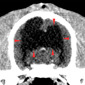
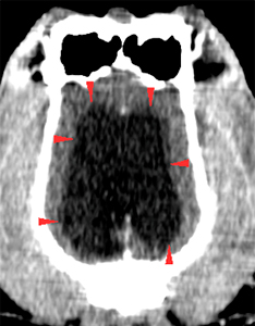
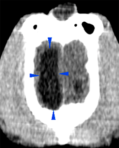
There is a moderate distention of the ventricular system, specifically of the central portions of the lateral ventricles, bilaterally, with absence of the “septum pellucidum”. This distention is more significant caudally, at the level of the occipital lobes (red arrowheads).
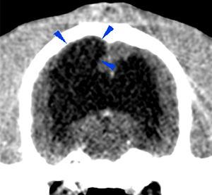
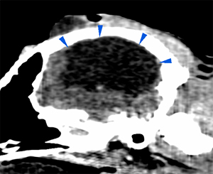
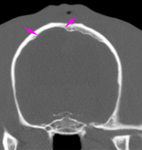
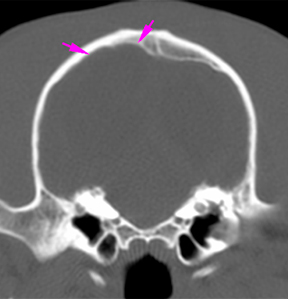
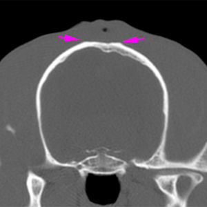
At the dorsal aspect of the right parieto-occipital region, there is an extension of the right lateral ventricle with complete loss of the cerebral cortex (dark blue arrowheads). At this level, the calvarium is slightly asymmetrical and deformed (pink arrows), compared to the contralateral side, with thinning of the bones in contact with the lesion described. However, there are no signs of bone discontinuity.
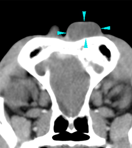
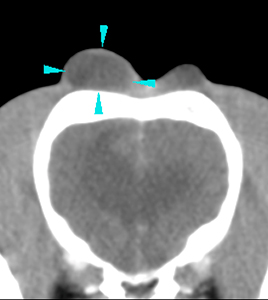
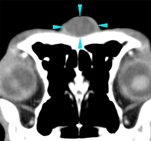
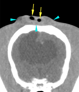
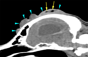
At the mid-dorsal aspect of the skull, there is a nodular thickening of the soft tissues, with round/oval shapes, hypoattenuating, without showing post-contrast enhancement. At least 4 rounded, well-defined, structures can be identified (blue arrowheads). Within some of these structures there are small gas bubbles (yellow arrows). Associated with the lesions located more caudally, there is a slight flattening of the outer surface of the parietal bone (pink arrows).
Diagnosis
Moderate/marked bilateral ventriculomegaly, with absence of the “septum pellucidum”, consistent with hydrocephalus of congenital origin, most likely.
Fluid attenuating lesion extending into the right parieto-occipital lobe, communicating with the right lateral ventricle, associated with a slight deformity of the calvarium, consistent with porencephaly most likely.
Multiple cystic lesions in the soft tissues of the mid-dorsal aspect of the skull. These have an uncertain origin and could be consistent with dermoid sinuses, considering their characteristics and other previously described intracranial congenitalabnormalities . Other differentials, such as granulomas, abscesses or hematomas, are considered less likely. A neoplastic process is considered unlikely. The gas bubbles could be iatrogenic due to previous sampling; concomitant infection (abscess with gas-producing bacteria vs. secondary to possible foreign body) is less likely.
Comments
In porencephaly, cystic cavities are present in the cerebrum due to cell destruction or failure of development. In this case, it is associated with hydrocephalus, as well as a slight malformation of the calvarium, which could explain the cognitive dysfunction presented by this patient.
It has been described that affected animals may be asymptomatic, show clinical signs related to the affected areas of the brain, including seizures, or, surprisingly, they could show neurological signs that are not normally located in the brain, such as nystagmus. Clinical signs may manifest later in the patient’s life. Correlation with the neurological exam and CSF tap are recommended.
On the other hand, the soft tissue lesions have an uncertain origin, although, considering their characteristics and location, multiple dermoid sinuses are considered the most likely differential. Dermoid sinus results from failure of separation of the surface ectoderm (forming the epidermis) and neuroectoderm (forming the nervous system) during embryogenesis. In this case, it could be a type V, considered a “true dermoid cyst”, with closed capsules and without deep connections with other tissues. FNA and analysis (cytology + culture), as well as histopathological analysis are recommended in order to reach a definitive diagnosis.
(Mon. to Fri. 9 a.m. to 6 p.m. gmt+1) Welcome, How can we help you?
We use cookies to ensure that we give you the best experience on our website. If you continue to use this site we will assume that you are happy with it.Ok


No comment yet, add your voice below!