12-year-old female Maltese. History of growing mass, ventral to the anus.
Description
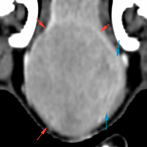
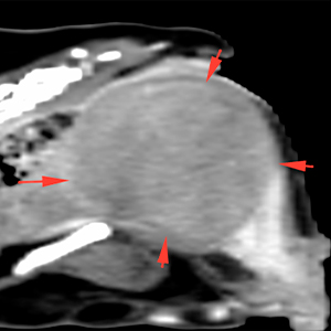
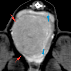
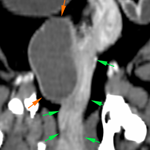
At intrapelvic level, and extending towards the perineal region, within the vagina, there is a round mass, with well-defined margins and soft tissue attenuation, showing a slightly heterogeneous contrast enhancement (red arrows). The mass appears to originate from the right wall of the vagina, growing eccentrically and markedly displacing the lumen of the vagina towards the left (blue arrows).
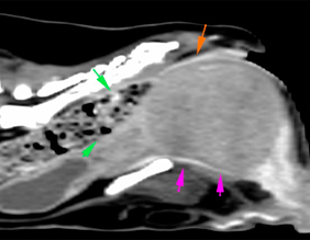
This mass causes a marked mass-effect, displacing the urethra ventrally (pink arrow), and the rectum and the anal region dorsally (orange arrows). Cranial to the mass, the colon shows a moderate accumulation of normal consistency faeces (green arrows).
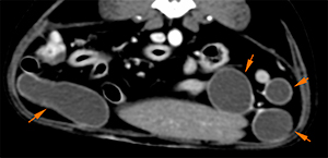
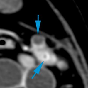
Cranial to the mass, the vagina and the uterine body are moderately thickened (green arrows). In the caudal abdomen, at the level of the uterine body, located on the right wall, there is a cystic lesion of eccentrical growth, with fluid attenuating content, surrounded by a thin capsule with increased contrast uptake (orange arrows).
Both uterine horns are moderately distended, filled with homogeneous fluid attenuating material, with a normal wall (orange arrows).
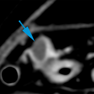
Both ovaries have a normal size and oval shape, with presence of small fluid attenuating lesions, showing well-defined margins (blue arrows).
Diagnosis
Vaginal mass, with mild intrapelvic and perineal extension, most likely consistent with neoplasia (leiomyoma, leiomyosarcoma).
Thickening of the uterine body with cystic structure of eccentric growth and moderate distension of both uterine horns with fluid content. Changes are consistent with cystic endometrial hyperplasia and hydrometra/mucometra/pyometra.
Small cysts vs. ovarian follicles.
comments
Mass in perianal/intrapelvic region, with vaginal origin, most likely neoplastic. Histopathological analysis is recommended in order to reach a definitive diagnosis. The other changes in the uterus are suggestive of cystic endometrial hyperplasia and hydrometra/mucometra/pyometra.
(Mon. to Fri. 9 a.m. to 6 p.m. gmt+1) Welcome, How can we help you?
We use cookies to ensure that we give you the best experience on our website. If you continue to use this site we will assume that you are happy with it.Ok


No comment yet, add your voice below!