Absence/aplasia of the vulva with atresia of the vestibular opening in a dog
10-months-old, neutered female Pinscher. Physical examination: she has neither penis nor vulva; however, the presence of a uterus was confirmed.
History of dysuria. An ovariohysterectomy was performed, and an incision was made in the urethral outlet to facilitate the passage of urine. Now she has dysuria again and accumulation of fluid around the stenosed hole of the urethra.
An abdominal CT scan was performed.
Description
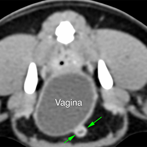
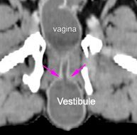
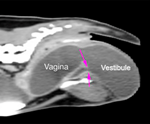
Absence of labia and vulva. There is a marked distension of the vagina and vestibule with hypoattenuating content and a thin hyperattenuating wall that enhances after contrast administration. These changes extend caudally, protruding and herniating between the soft tissues of the perineal area, with the vestibule appearing at subcutaneous level (orange arrows). There is a discreet irregularity on the surface of the most caudoventral aspect of the vestibule, without any obvious opening to the outside. The distention produces a mass-effect with compression of the rectum dorsally and of the urethra ventrally.
The urinary bladder is moderately distended, with hypoattenuating content and homogeneous wall. The urethra is completely visible, showing a slightly thickened wall that enhances after contrast administration (green arrows). The urethrovestibular junction/urethral opening has a normal appearance, located dorsal to the caudal aspect of the pubis and 4.5 cm from the most caudal aspect of the vestibule, adjacent to the skin (pink arrows).
Diagnosis
Absence/aplasia of the vulva with atresia of the vestibular opening towards the outside, consistent with a congenital malformation.
Vestibule-vaginal distension, possibly filled with urine due to obstruction and stenosis at the level of the opening, secondary to the previous surgery.
Comments
In this case, it is complicated to differentiate between pre- and post-surgical alterations. However, there seems to be a malformation at the level of the caudal urogenital tract, and stenosis of the previously performed opening.
(Mon. to Fri. 9 a.m. to 6 p.m. gmt+1) Welcome, How can we help you?
We use cookies to ensure that we give you the best experience on our website. If you continue to use this site we will assume that you are happy with it.Ok


No comment yet, add your voice below!