8-year-old female neutered crossbreed. History of progressive loss of mobility in the hindlimbs. Came to the emergency room with paraparesis and loss of deep pain perception.
A thoracolumbar spine and thoracic CT scan was performed.
Description
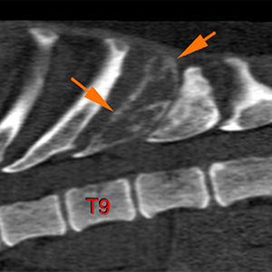
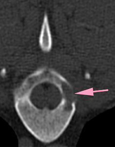
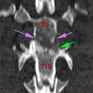
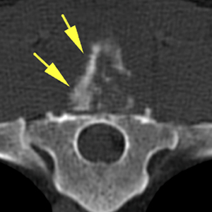
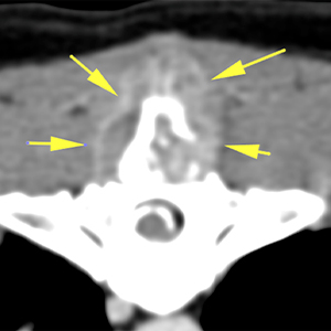
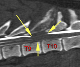
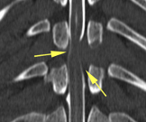
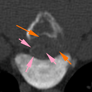
There are 13 thoracic vertebrae, with T13 showing 2 normal ribs. There is an expansile and destructive lesion affecting the spinous process (orange arrows), left pedicle (pink arrow), dorsal lamina and both caudal articular processes of T9 (purple arrows), as well as the left cranial articular process of T10 (green arrow). This lesion shows an osteolytic permeative pattern of the medullary cavity, with disruption of the cortices and a spiculated periosteal reaction (yellow arrows), especially at the spinous process, with an ill-defined transition area.
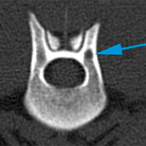
In the post-contrast study, performed after the MyeloCT, the lesion shows a soft tissue component that is more clearly visible, showing a marked and heterogeneous contrast enhancement, affecting the epaxial musculature, surrounding the spinous process of T9 (yellow arrows) and infiltrating the vertebral canal on the left and ventrally, as well as the left intervertebral foramen (blue arrows) which is slightly widened. This lesion displaces and compresses the spinal cord.
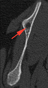
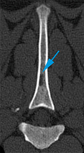
On the spine of the right scapula, there is a round, geographical lytic lesion, that does not affect the cortex, with a short transitional zone (well defined) (red arrow). There are a couple of similar lesions affecting the spinous process of T3 and left pedicle of T13 (blue arrows).
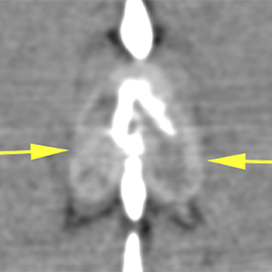
MyeloCT (contrast injection at L5-L6): The dorsal and ventral contrast columns can be followed correctly until the cranial aspect of T10, where they are attenuated and displaced towards the right and ventrally at the level of the vertebral body of T9 (yellow arrows) by an extradural lesion, located dorsally and to the left (lesion previously described) (orange arrows). This lesion displaces the spinal cord towards the right, compressing it moderately (pink arrows). Cranial to T9, the contrast columns recover a normal appearance.
Diagnosis
Expansile and aggressive osseous lesion affecting T9 and the cranial articular process of T10, with a soft tissue component that extends into the vertebral canal, consistent with a neoplastic process (osteosarcoma, most likely; chondrosarcoma or fibrosarcoma, less likely). The lesion behaves like an extradural lesion, causing a moderate/severe compressive myelopathy based on the MyeloCT findings.
Geographic lytic lesions in the right scapula, spinous process of T3 and left pedicle of T13, can be consistent with degenerative changes (cysts), although bone metastasis could also be possible, taking into account the remaining findings.
Thorax without evident abnormalities.
Comments
CT guided or ultrasound guided FNAs of the lesion at T9 is recommended in order to reach a definitive diagnosis. There are no signs of metastasis in the pulmonary parenchyma
(Mon. to Fri. 9 a.m. to 6 p.m. gmt+1) Welcome, How can we help you?
We use cookies to ensure that we give you the best experience on our website. If you continue to use this site we will assume that you are happy with it.Ok


No comment yet, add your voice below!