Aortic thromboembolism and left renal ischemia in a dog
9-years-old crossbreed dog with ambulatory paraparesis. A CT-scan of the thoracic and lumbosacral spine was performed.
Description
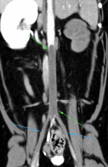
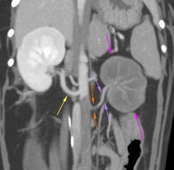
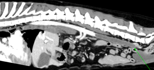
There is a large and elongated filling defect in the caudal abdominal aorta (green arrows), that runs from the cranial aspect of the left renal artery through the external iliac arteries up to the femoral arteries (blue arrows). This lesion causes a complete obliteration of the lumen of the aorta and the iliac arteries. The left renal artery is smaller in size compared to the right (yellow arrow) and shows a homogeneous contrast enhancement in its distal portion (purple arrows). A small filling defect is seen in the proximal aspect of this vessel (orange arrows).
The left kidney is irregular and mildly diminished in size with no contrast enhancement (pink arrows), only in some small arterial vessels. No contrast enhancement is seen along the left renal vein.
The right kidney and the rest of the urinary tract do not show abnormalities.
The thoracic and lumbar spine included in the study does not show significant abnormalities.
Diagnosis
Severe aortic thromboembolism from the level of the left renal artery up to the region of the femoral arteries
Changes in the left kidney consistent with renal ischemia.
Comments
The thrombosis could be caused by a hypercoagulable state due to heart disease, bacterial infection (sepsis or septicemia), neoplasia, injury of a blood vessel’s lining, parasitic heart disease, hyperadrenocorticism (with no adrenal gland lesion identified on the CT-scan), hyperthyroidism, platelet abnormalities or protein-losing nephropathy, among others. Echocardiography is recommended to assess cardiac functionality, as well as in order to rule out systemic alterations that may have caused the thromboembolism.
(Mon. to Fri. 9 a.m. to 6 p.m. gmt+1) Welcome, How can we help you?
We use cookies to ensure that we give you the best experience on our website. If you continue to use this site we will assume that you are happy with it.Ok


No comment yet, add your voice below!