6-year-old male dog. History of chronic cough.
A thoracic CT scan was performed.
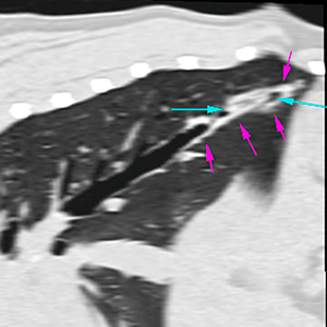
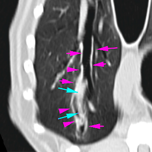
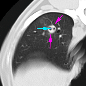
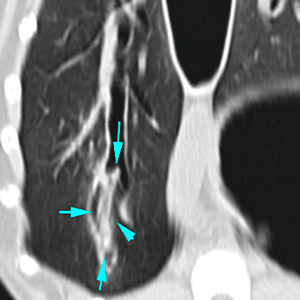
Description
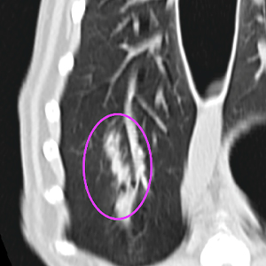
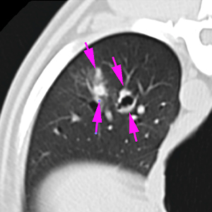
The lung parenchyma is well expanded, without signs of nodular lesions or masses. There is a moderate / marked regional luminal distention with circumferential thickening of the bronchial wall affecting the distal aspect of the lobar bronchi of the right caudal lung lobe (pink arrows). In addition, in this area, there is an intraluminal structure, with ill-defined margins, soft tissue attenuation and longitudinal appearance (approx. 3 cm in length) (blue arrows). These changes are associated with a soft tissue attenuating tubular area, with peribronchial distribution, that extends cranially from the bronchial changes previously described (purple arrows and circle). Mild peripheral increase in attenuation (ground-glass) in this region.
Diagnosis
Presence of an inhaled foreign body (plant material / grass awn) located in the distal aspect of the lobar bronchi of the right caudal lung lobe, with associated mild focal bronchopneumonia.
comments
There is an intraluminal structure visible in the lobar bronchi of the right caudal lung lobe, that suggests a plant / grass awn-type bronchial foreign body associated with focal bronchopneumonia, which justifies the clinical signs of the patient.
Consider bronchoscopy in order to remove the foreign body.
(Mon. to Fri. 9 a.m. to 6 p.m. gmt+1) Welcome, How can we help you?
We use cookies to ensure that we give you the best experience on our website. If you continue to use this site we will assume that you are happy with it.Ok


No comment yet, add your voice below!