2-year-old male crossbreed dog, rescued with left forelimb lameness, decreased extension and flexion of the left shoulder and soft tissue swelling.
A shoulder CT-scan was performed.
Description
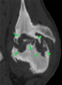
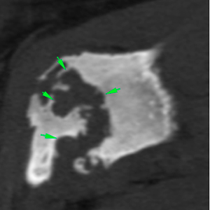
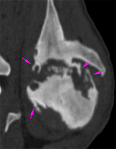
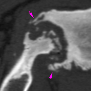
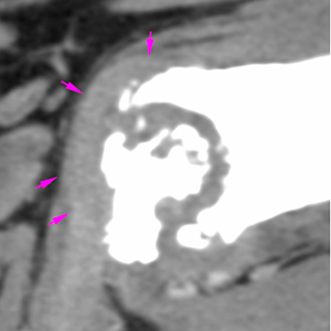
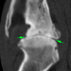
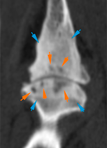
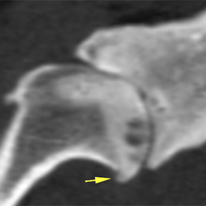
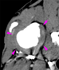
Left shoulder
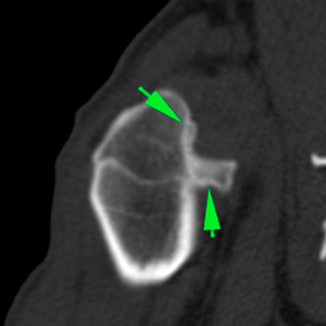
There are severe bone lesions at the left shoulder. There are multiple areas of moth-eaten and geographic bone lysis, affecting the greater tubercle and the head of the humerus, as well as the glenoid cavity of the scapula (green arrows). In addition, there is an extensive subchondral bone destruction with an irregular articular surface (orange arrows). There is marked smooth and irregular periarticular new bone formation and areas of sclerosis at the distal scapula and proximal humerus (yellow arrows). These changes cause a marked deformity of the scapulohumeral joint (pink arrows).
There is a severe thickening of the periarticular soft tissues, showing heterogeneous contrast enhancement (orange arrows). No joint effusion is detected.
The tendon of the biceps muscle is not clearly visible. However, in some areas it appears to be slightly thickened (pink arrows).
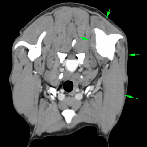
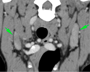
There is moderate/marked atrophy of the musculature of the left forelimb (green arrows).
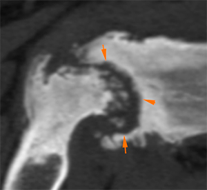
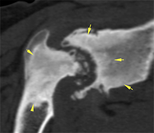
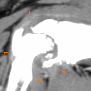
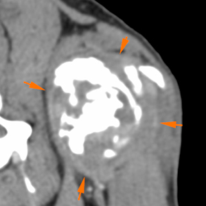
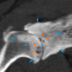
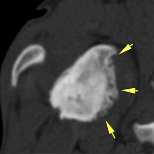
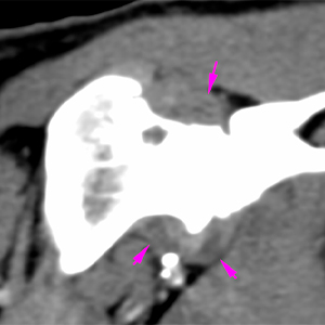
Right shoulder
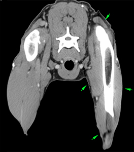
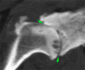
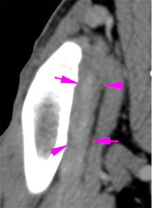
The changes visible at the right shoulder are similar as the ones previously described at the left shoulder, but less severe. There is a severe collapse of the joint space (green arrows). In addition, there are multiple moth-eaten and geographic lytic bone lesions at the proximal humerus and distal scapula affecting the subchondral bone (orange arrows). These lesions are surrounded by areas of sclerosis (blue arrows).
There is moderate periarticular new bone formation at the humeral head and along the medial aspect of the scapula (yellow arrows). There is also a moderate/marked new bone formation at the intertubercular groove surrounding the biceps tendon (green arrows). The tendon is thickened, surrounded by a hypoattenuating halo consistent with a distended sheath (pink arrows).
There is periarticular soft tissue swelling, as well as distention of the joint capsule and a moderate joint effusion (pink arrows).
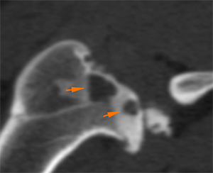
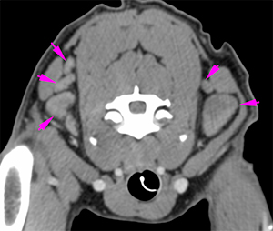
The cervical superficial and axillary lymph nodes are markedly enlarged (left > right) – pink and green arrows respectively -.
Diagnosis
– The findings in both shoulders are consistent with chronic erosive joint disease and moderate bone remodeling. The differentials include previous/old septic arthritis vs. active infection (bacterial or fungal less likely with haematogenous spread), other infectious causes such as Leishmania, or immune-mediated polyarthritis (rheumatoid arthritis) could be possible. Other causes such as chronic haemarthrosis are less likely. Associated findings:
– Marked collapse of the right scapulohumeral joint and incongruity of the left.
– Bilateral periarticular soft tissue swelling with moderate effusion in the right shoulder.
– Findings at both biceps muscle tendons: most likely consistent with associated bilateral bicipital tendinopathy (R>L).
Marked bilateral cervical superficial and axillary lymphadenopathy: reactive (infectious vs immune-mediated process).
Comments
Arthrocentesis of both scapulohumeral joints with complete analysis, including culture, of the synovial fluid are recommended for further investigations.
(Mon. to Fri. 9 a.m. to 6 p.m. gmt+1) Welcome, How can we help you?
We use cookies to ensure that we give you the best experience on our website. If you continue to use this site we will assume that you are happy with it.Ok


No comment yet, add your voice below!