12-year-old male cat, suffering neurological symptoms, such as circling and left head tilt, with sudden blindness and behaviour changes. A head CT scan was performed.
REPORT
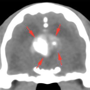
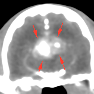
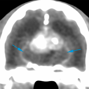
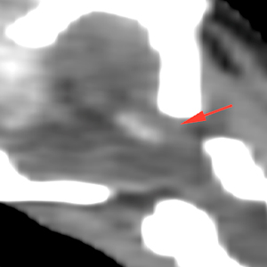
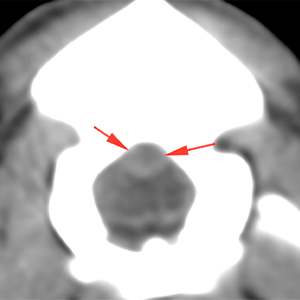
Intracranially, there are 2 extra-axial lesions (red arrows) that seem to originate from both lateral ventricles, extending caudally, dorsal to the mesencephalon. Both lesions have well-defined and irregular margins, are hyperattenuating (140 HU), with mineralized material inside (420 HU), showing a moderate post-contrast enhancement.
Pre-contrast
Post-contrast
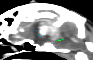
These lesions cause a marked mass effect, displacing and compressing the diencephalon and mesencephalon ventrally (blue and green arrows respectively).
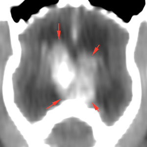
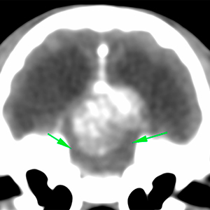
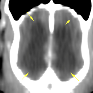
There is a marked, bilateral and symmetrical, distention of both lateral ventricles (yellow arrows). There is also a mild / moderate cerebellar herniation, with cerebellar extension through the foramen magnum (red arrows).
Diagnosis
Mineralized extra-axial lesions in both lateral ventricles, consistent with cholesterol granulomas, most likely, considering their characteristics. Other differentials, such as a neoplastic disease (e.g. choroid plexus tumour) are less likely, although cannot be ruled out.
Associated obstructive hydrocephalus and mild cerebellar herniation.
(Mon. to Fri. 9 a.m. to 6 p.m. gmt+1) Welcome, How can we help you?
We use cookies to ensure that we give you the best experience on our website. If you continue to use this site we will assume that you are happy with it.Ok

No comment yet, add your voice below!