2.5 months-old, male Spanish Water Dog. Previous history of parvovirus. Now he shows lumbar and joint pain. Physical examination: ambulatory paraparesis and fever.
A spinal, fore limbs and pelvic limbs CT scan was performed.
Description
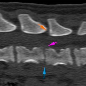
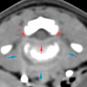
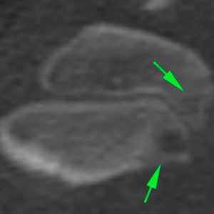
Spine
There is a collapse of the intervertebral disc space of L5-L6 (blue arrow), with a slight ventral displacement of L6. There is a small step of approx. 2 mm between the vertebral bodies of L5-L6 (pink arrow) and the articular processes of both vertebrae (orange arrow).
The caudal and cranial endplates of L5 and L6, respectively, are slightly irregular, with moth-eaten areas of lysis (yellow arrows), as well as mild adjacent sclerotic changes (green arrows), and a slightly irregular periosteal reaction along the ventral aspect of both vertebral bodies (orange arrows).
The soft tissues of the intervertebral disc space of L5-L6 are thickened, irregular and with strong contrast enhancement, affecting the hypaxial muscles (blue arrows) and extending ventrally along the vertebral canal (red arrows), causing a loss of visualization of the epidural fat and a minimal dorsal displacement of the spinal cord, without signs of compression or other obvious intramedullary lesions.
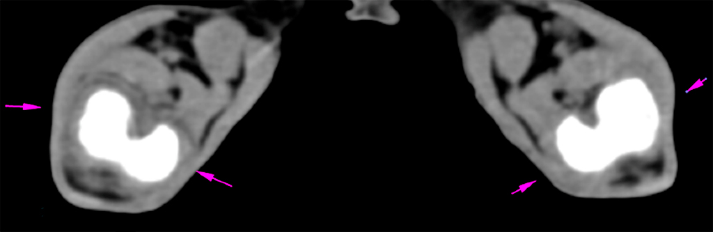
Hind limbs
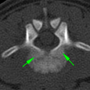
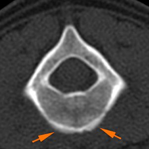
There is a moderate joint effusion in both stifles, being slightly more extensive in the right stifle (orange arrows). The periarticular soft tissues are thickened and hyperattenuating (pink arrows), being more severe in the right stifle. There are no evident osseous lesions.
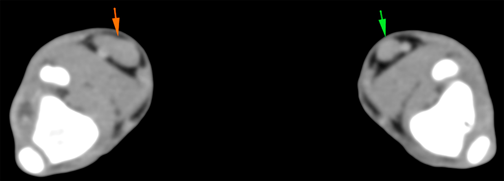
The right popliteal lymph node (orange arrow) is slightly more prominent and asymmetric in comparison to the contralateral side (green arrow).
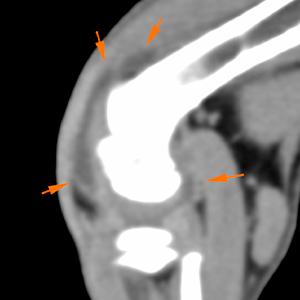
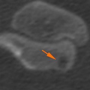
Fore limbs
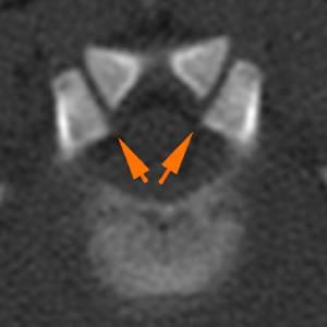
There is a mild joint effusion in both shoulders (green arrows), more severe in the left shoulder, with moderate thickening of the periarticular soft tissues, which are strongly enhancing after contrast administration (yellow arrows).
In the left shoulder, at the level of the caudal aspect of the humeral head and in the region of the proximal metaphysis, there are small osteolytic lesions with slightly irregular margins (green arrows). In the right shoulder, there is another lytic lesion in the area of the proximal metaphysis of the humerus (orange arrow).
Diagnosis
Collapse of the intervertebral disc L5-L6, with lysis of the endplates of both vertebrae, mild ventral periosteal reaction and post-contrast enhancement of the hypaxial musculature and soft tissues at the ventral aspect of the vertebral canal. This is consistent with an inflammatory/infectious process (acute discospondylitis, most likely). The lesion is causing a mild subluxation of the spine at this level.
Thickening in periarticular soft tissues of shoulders and stifles, with associated joint effusion and osteolytic lesions in the proximal aspect of both humerus (with more severe changes in the left). The findings are consistent with an inflammatory/infectious process (septic polyarthritis, most likely).
Mild right popliteal lymphadenopathy most likely reactive.
Comments
The changes described are most likely consistent with an inflammatory/infectious process probably due to septicaemia, that has spread to the spine and limbs (discospondylitis and polyarthritis).
Sampling (arthrocentesis, blood culture, urine culture) is required to reach a definitive diagnosis. Pseudomonas aeruginosa was isolated in the blood culture.
(Mon. to Fri. 9 a.m. to 6 p.m. gmt+1) Welcome, How can we help you?
We use cookies to ensure that we give you the best experience on our website. If you continue to use this site we will assume that you are happy with it.Ok


No comment yet, add your voice below!