2-year-old female, crossbreed dog. She has been avoiding any kind of activity for 4-5 weeks. Then she started with lameness of the right pelvic limb.
A thoracolumbar spine and thoracic CT scan was performed.
Description
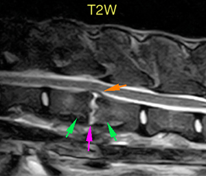
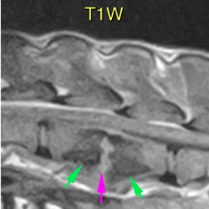
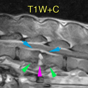
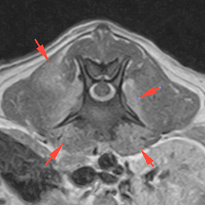
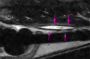
There is a narrowing/collapse of the intervertebral disc space of L2-L3, that shows an abnormal shape. The normal disc has been replaced by a hyperintense heterogeneous signal on T2W and STIR, showing homogeneous contrast enhancement (pink arrows). The vertebral endplates are irregular, with hypointense signal on T2W and T1W compared to the normal bone marrow, consistent with sclerosis (green arrows).
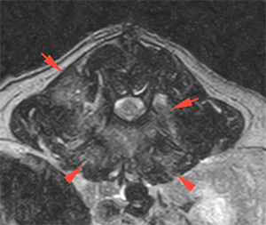
At this location, in the vertebral canal, there is an attenuation of the ventral subarachnoid space on T2W (orange arrow), with mild stenosis of the vertebral canal and both intervertebral foramina, with enhancement of this region on the post-contrast sequence (blue arrows). This causes a mild displacement and compression of the spinal cord.
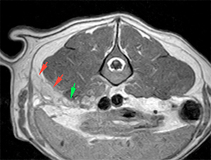
Adjacent to this region, there is a diffuse hyperintensity on T2W and STIR of the paravertebral muscles (hypaxial and epaxial), on both sides, more evident on the right, showing homogeneous contrast enhancement (red arrows).
T2W
T1W+C
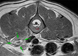
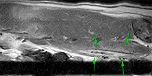
There is moderate thickening of the right psoas muscle. In this area, there is a cavitary lesion with a fusiform shape, with hypointense material at its core, with a hyperintensity halo (fluid) on T2W surrounding it (pink arrows). After contrast administration, the lesion shows peripheral enhancement (green arrows). This lesion extends from the cranial aspect of L5 to the caudal aspect of L6.
T2W
T1W+C
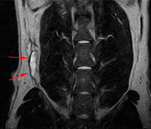
At the level of L3-L4, there is a collection of hyperintense material on T2W, with mixed signal intensity on T1W and showing a slightly heterogeneous contrast enhancement, located in the subcutaneous tissue of the caudal aspect of the right abdominal wall, cranial to the iliac wing (red arrows). There is a fistulous tract connecting this lesion with the previously described lesion in the psoas muscle (green arrow).
T2W
T1W+C
Diagnosis
Discospondylitis at the intervertebral space of L2-L3, with extension of the inflammation/infection into the paravertebral musculature, as well as ventral epidural region and intervertebral foramina. This causes a mild stenosis of the vertebral canal and mild ventral compression of the spinal cord and nerve roots at this location.
Lesion at the level of the right psoas muscle, consistent with an abscess (probably associated to a migratory foreign body, i.e., grass awn, splinter, etc.) most likely, with associated fistulous tract connecting with a subcutaneous abscess at the right abdominal wall.
Comments
Consider US assessment of the right psoas muscle in the region of L5-L6 to try to visualize the foreign body and US-guided drainage of the subcutaneous abscess at the abdominal wall.
(Mon. to Fri. 9 a.m. to 6 p.m. gmt+1) Welcome, How can we help you?
We use cookies to ensure that we give you the best experience on our website. If you continue to use this site we will assume that you are happy with it.Ok


No comment yet, add your voice below!