12-year-old male Maltese with respiratory distress that worsened within hours. Thoracic CT was performed.
A thoracolumbar spine and thoracic CT scan was performed.
Description
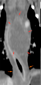
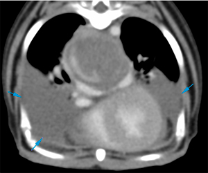
There is an esophageal mass growing eccentrically from the left oesophageal wall (red arrows) with diffuse and circumferential thickening of the wall of the caudal oesophagus (orange arrows). The mass causes displacement and compression of the oesophageal lumen to the right (blue arrows). The mass shows slightly heterogeneous soft tissue attenuation, with mild peripheral contrast enhancement
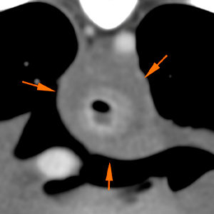
It causes a moderate mass effect by displacing the carina and heart ventrally (green arrows), as well as a moderate narrowing of the carina and both main stem bronchi (red arrows).
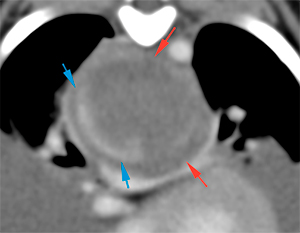
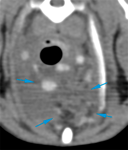
In the cranioventral mediastinum, there is a moderate thickening with diffuse increased attenuation of the mediastinal fat (blue arrows).
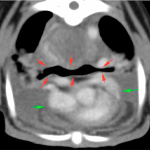
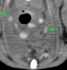
The mediastinal lymph nodes are slightly prominent, showing a marked and homogeneous post-contrast enhancement (green arrows).
There is a moderate amount of pleural effusion in the dependent part of both hemithorax (blue arrows).
Diagnosis
Esophageal mass at the level of the caudal thoracic esophagus consistent with neoplasia (leiomyoma or leiomyosarcoma, most likely rather than another type of tumor considering its eccentric growth). Other ddx such as a granulomatous lesion are unlikely. This mass causes narrowing of the carina and main stem bronchi.
Thickening of the mediastinum: mediastinitis or neoplastic infiltration.
Mild mediastinal lymphadenopathy: reactive or metastatic.
Moderate bilateral pleural effusion.
Comments
The findings are consistent with an oesophageal mass, most likely consistent with a neoplastic process. This mass causes moderate compression of the carina and both main stem bronchi, in addition to moderate pleural effusion, which could explain the patient’s respiratory symptoms described in the history. Histopathological analysis of the mass is required to reach a definitive diagnosis, as well as analysis of the pleural effusion.
(Mon. to Fri. 9 a.m. to 6 p.m. gmt+1) Welcome, How can we help you?
We use cookies to ensure that we give you the best experience on our website. If you continue to use this site we will assume that you are happy with it.Ok


No comment yet, add your voice below!