8-year-old female Jack russel terrier with cervical pain and ataxia.
An MRI of the head was performed.
Description
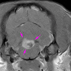
There is an intracranial and extra-axial mass located rostral to the fourth ventricle. It is hyperintense on T2W and FLAIR, hypointense on T1W, showing a strong and slightly heterogeneous contrast enhancement with a small intralesional hypointense area (pink arrows).
T1W+C
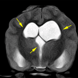
T2W
T1W+C
The lesion produces a moderate mass effect with displacement and compression of the brainstem ventrally and of the cerebellum caudodorsally in the caudal fossa. There is focal loss of cerebrospinal fluid signal (CSF) at the level of the foramen magnum with slight protrusion of the caudal aspect of the cerebellum.
Rostral to the previously described lesion, the lateral ventricles (slightly asymmetric R>L) and third ventricle are dilated (yellow arrows). There is also a moderate supracolicular fluid accumulation.
T2W
At the level of the cervical spine, the central canal is dilated with a diffusely hyperintense signal on T2W/FLAIR, hypointense on T1W, with no obvious contrast enhancement (orange arrows).
T2W
Diagnosis
Extra-axial mass in the area of the fourth ventricle. This mass causes mild obstructive hydrocephalus, cerebellar herniation secondary to mass effect and dilatation of the central canal at the cervical spine (syringomyelia). These findings are consistent with neoplasia – choroid plexus tumor (papilloma, carcinoma) vs ependimoma or meningioma.
(Mon. to Fri. 9 a.m. to 6 p.m. gmt+1) Welcome, How can we help you?
We use cookies to ensure that we give you the best experience on our website. If you continue to use this site we will assume that you are happy with it.Ok


No comment yet, add your voice below!