11-year-old male neutered miniature Schnauzer. Extrahepatic portosystemic shunt (PSS) is suspected on ultrasound. An abdominal CT scan was performed.
Description
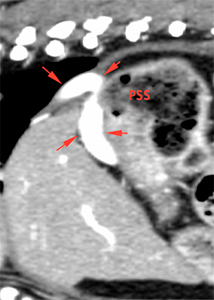
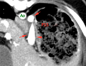
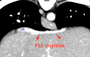
There is a large abnormal vessel (red arrows, PSS), originating from the left gastric vein. This vessel runs craniodorsally, parallel to the lesser curvature of the stomach, and at the level of the diaphragm, is located ventral and to the left of the aorta. At this level, it turns to the right, running cranial to the liver, inserting into the caudal vena cava through the phrenic vein in the region of confluence of the hepatic veins
Cranial to the origin of the shunt, the portal vein shows a slight reduction in diameter (green line) compared with its diameter caudal to the shunt (red line), gradually increasing in size again when it receives its last tributary (gastroduodenal vein). There is adequate intrahepatic portal ramification (orange arrows).
Hepatic and renal size are within the normal limits.
The gallbladder is moderately distended with small, round, mineral attenuating structures deposited in the dependent lumen (orange arrows). No signs of obstruction are noted.
Diagnosis
Left gastric-phrenic shunt (extrahepatic portosystemic shunt),of congenital origin, most likely.
Portal vein with a correct diameter at the level of the porta hepatis and normal intrahepatic portal vascularization.
Small choleliths without signs of obstruction
No changes in liver or kidneys are observed.
Comments
Miniature schnauzers may not necessarily show signs of hepatic insufficiency as obviously as other breeds, and these dogs experienced less hepatic insufficiency than other dogs with congenital PSS. For this reason, it is sometimes diagnosed in adult animals. In addition, it has been reported that spleno-phrenic shunts are diagnosed at a significantly older age than other types of shunts. The phrenic vein drains into the CVC caudal to the diaphragm and cranial to the liver, and readily collapses with each respiratory cycle. This could contribute to decreased shunt fraction and result in latent clinical signs and preserver portal vein size.
(Mon. to Fri. 9 a.m. to 6 p.m. gmt+1) Welcome, How can we help you?
We use cookies to ensure that we give you the best experience on our website. If you continue to use this site we will assume that you are happy with it.Ok


No comment yet, add your voice below!