A 1-year-old, female, spayed crossbreed dog was presented with fever and abdominal pain. In the abdominal ultrasound, pelvic and ureteral distention were seen.
An abdominal CT was performed.
Description
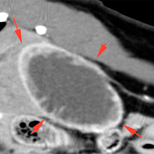
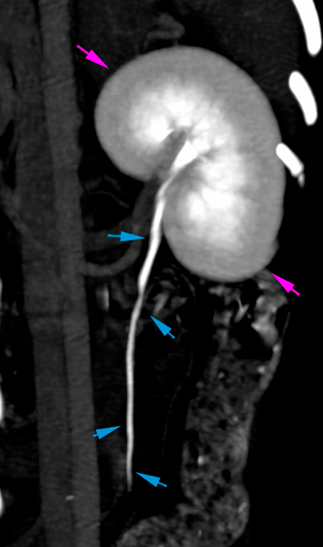
The right kidney is mildly increased in size compared to the left kidney. The right renal pelvis is markedly distended. There is loss of the normal renal architecture, as well as a marked thinning of the renal cortex, with multiple septa showing post-contrast enhancement. The central portion of the right kidney is filled with fluid attenuating, non-enhancing content consistent with marked hydronephrosis (red arrows).
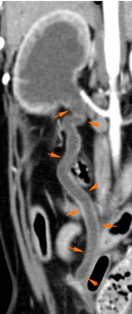
The right ureter is markedly and diffusely distended (up to 9 mm) with fluid content (orange arrows). The ureter’s wall does not show abnormalities with normal homogeneous enhancement
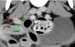
At the level of L7, the right ureter is markedly narrowed (green arrows). In the retroperitoneum, adjacent to the ureter, there is a nodular soft tissue lesion that shows a marked and homogeneous post-contrast enhancement. This lesion also extends towards the right psoas muscle (red arrows).
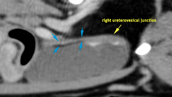
Caudal to this lesion, the ureter shows a normal size (2 mm) (blue arrows) and can be seen up to the right ureterovesical junction (yellow arrows).
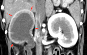
The left kidney has normal size, shape and renal architecture, without distention of its pelvis (pink arrows). The left ureter is unremarkable, normally filled with contrast along its path (blue arrows).
Diagnosis
Right hydronephrosis and hydroureter, secondary to obstruction of the right ureter.
Lesion in the distal aspect of right ureter: ureteral stenosis with peritoneal nodular lesion that extends to the right psoas muscle, consistent with inflammatory/granulomatous lesion, most likely. Ddx include: granuloma or adhesions related to previous surgery (ovariohysterectomy) most likely, inflammation of the ureter – secondary to urinary tract infection – or related to trauma.
Left kidney and ureter within normal limits.
Comments
The findings in the right ureter are consistent with a region of ureteral stricture causing right hydroureter and hydronephrosis. Considering the lesion in the retroperitoneum and enhancement of the right psoas muscle, the presence of a granulomatous lesion, adhesions secondary to previous surgery or inflammation should be considered as the most likely causes of ureteral stricture.
(Mon. to Fri. 9 a.m. to 6 p.m. gmt+1) Welcome, How can we help you?
We use cookies to ensure that we give you the best experience on our website. If you continue to use this site we will assume that you are happy with it.Ok


No comment yet, add your voice below!