Intestinal mass with origin in the cecum and septic peritonitis in a dog
10-year-old male Maltese with abdominal pain.
An abdominal CT scan was performed.
Description
Abdomen
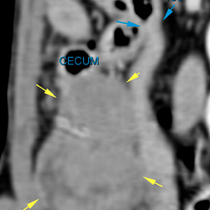
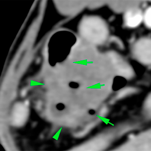
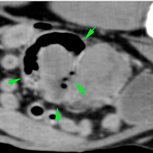
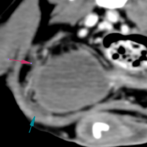
In the right middle abdomen, located immediately caudal to the ileocolic junction, there is a well-defined mass, with slightly irregular margins, soft tissue heterogeneous attenuation, showing a marked and heterogeneous contrast enhancement (yellow arrows). The mass is in intimate contact with part of the serosa of the ascending colon and cecum, part of which seems to be embedded inside the mass affecting its wall (green arrows), containing fluid and gas inside the lumen. The ileocolic junction does not show any abnormalities (blue arrows).
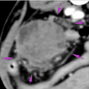
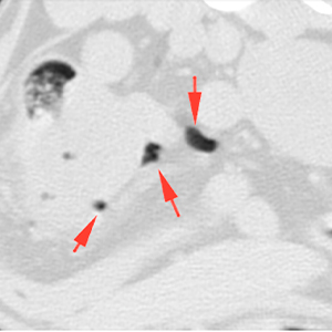
Around the mass, the peritoneum is heterogeneous and hyperattenuating “fat stranding” (purple arrows). There is a small amount of free fluid in its depending portion, ventral to the bladder (blue arrow), as well as several gas bubbles (red arrows).
There are no signs of intra-abdominal lymphadenopathy, including jejunal or right colic lymph nodes adjacent to the previously described mass.
Diagnosis
Abdominal mass with an intestinal origin, most likely, originating from the cecum. These findings are consistent with neoplastic disease: gastrointestinal stromal tumor (GIST) or leiomyosarcoma, most likely; or carcinoma, less likely. Other differentials, such as granuloma, are less likely.
Heterogeneous peritoneum with mild peritoneal effusion and mild pneumoperitoneum, consistent with septic peritonitis most likely, associated to intestinal perforation.
NOTE: In this case the Gastrointestinal Stromal Tumor was confirmed by histopathological analysis after surgery.
(Mon. to Fri. 9 a.m. to 6 p.m. gmt+1) Welcome, How can we help you?
We use cookies to ensure that we give you the best experience on our website. If you continue to use this site we will assume that you are happy with it.Ok


No comment yet, add your voice below!