2-years-old, male, Pointer presented with acute and progressive paraparesis for 5 days. An MRI of the thoracolumbar spine was performed.
Description
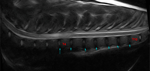
The spine is included from C6 to L4, showing a normal alignment. The vertebral count is normal with 13 thoracic vertebrae and T13 has a pair of symmetrical ribs. The celiac trunk is located ventral to L1.
There are no aggressive bone lesions at the vertebrae, intervertebral space or adjacent soft tissue.
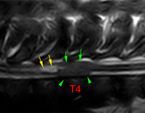
There is loss of the normal T2W hyperintense signal of the nucleus pulposus at the intervertebral discs from T3 to T10 (blue arrows).
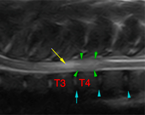
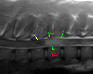
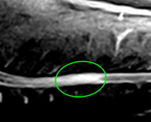
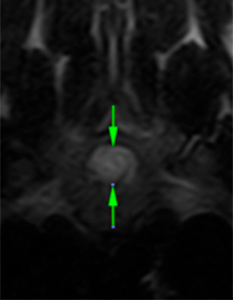
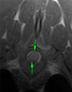
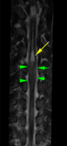
There is a focal rounded intramedullary lesion at the level of T4, resulting in peripheral thinning of the normal T2W hyperintense signal of the cerebrospinal fluid and epidural fat (green arrows). The lesion is mildly hyperintense compared to the spinal cord on T2W, STIR (green circle) and T1W+Gd. No marked meningeal enhancement or signal voids are seen on T2*. There is no associated extradural component.
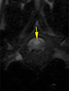
The central canal is mildly dilated at the caudal aspect of T3, immediately cranial to the lesion described, hyperintense on T2W, and hypointense on T1W suggesting cerebrospinal fluid (yellow arrows).
T2W
T1W+Gd
STIR
T2W
T1W+Gd
STIR
T2W
T1W+Gd
Diagnosis
Intramedullary lesion with mass effect at the level of the T4, most likely consistent with intramedullary neoplasia – glioma, ependymoma, round cell tumor -, infectious or inflammatory meningomyelitis or ischemic myelopathy are less likely.
Focal dilation of the central canal, consistent with syringohydromyelia secondary to the described intramedullary lesion.
Chronic intervertebral disc degeneration T3-T10.
Comments
The clinical sings are probably related to the intramedullary lesion at the level of T4. Cerebrospinal fluid tap and analysis are recommended for further investigations.
(Mon. to Fri. 9 a.m. to 6 p.m. gmt+1) Welcome, How can we help you?
We use cookies to ensure that we give you the best experience on our website. If you continue to use this site we will assume that you are happy with it.Ok


No comment yet, add your voice below!