7-year-old, female neutered, Weimaraner presented with a hyperacute-onset monoparesis of the right hindlimb. No obvious signs of hyperesthesia. Neurolocalisation: Lumbar intumescence.
Description
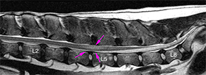
There are 8 lumbar vertebrae. The intervertebral (IV) discs of T13-L1 and L2-L4 are dehydrated with loss of the normal hyperintense signal in T2W.
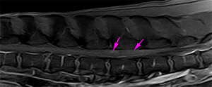
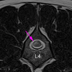
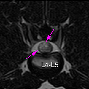
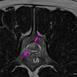
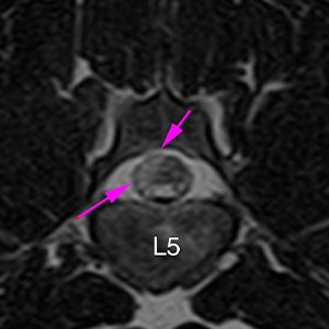
There is an intramedullary lesion, with ill-defined margins, located at the right lateral aspect of the spinal cord at the level of L4-L5 (lumbar intumescence). These changes extend from the mid L4 to the caudal aspect of L5, occupying a slightly larger medullary diameter at the IV space of L4-L5. The lesion is hyperintense in T2W, isointense in T1W and does not show a susceptibility artifact in T2* (pink arrows), showing a mild contrast enhancement. There is no spinal cord compression at this level.
There is a mild hypertrophy of the dorsal longitudinal ligament at the level of T13-L4 and L7-S1, with a decreased signal of the ventral epidural fat, without evidence of spinal cord compression.
Diagnosis
Intramedullary lesion lateralized to the right at L4-L5, consistent with ischemic myelopathy (fibrocartilaginous embolism).
(Mon. to Fri. 9 a.m. to 6 p.m. gmt+1) Welcome, How can we help you?
We use cookies to ensure that we give you the best experience on our website. If you continue to use this site we will assume that you are happy with it.Ok


No comment yet, add your voice below!