1-year-old male German Shorthaired Pointer. The ophthalmological exam revealed exophthalmia, mydriasis and absence of pupillary reflex. There is no response to medical treatment during 2 weeks.
MRI of the head was performed.
DESCRIPTION
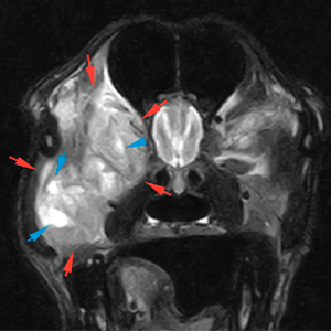
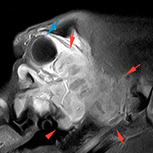
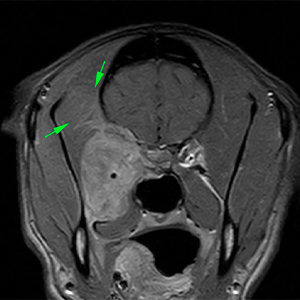
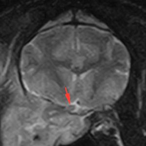
At the level of the right orbit, mainly at its lateroventral aspect, there is a large mass with irregular and well-defined margins (red arrows). The mass is hyperintense on T2W and isointense on T1W, in comparison to the musculature, showing marked and slightly heterogeneous contrast enhancement, with presence of multiple small cavities hyperintense on T2W and hypointense on T1W, without contrast enhancement (blue arrows).
T2W
T1W
T1W+Gd
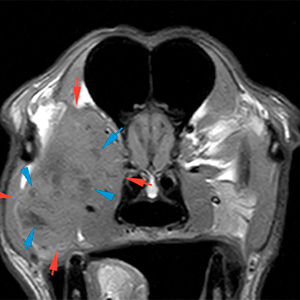
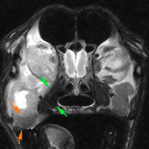
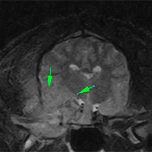
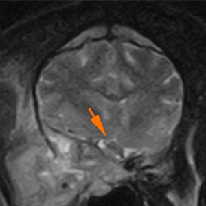
The mass causes a marked mass-effect, causing a rostrodorsal displacement of the ocular globe (exophthalmos) (blue arrow), which seems to be intact, and a ventral displacement of the zygomatic gland (orange arrows) and the medial pterygoid muscle (green arrows).
T1W+Gd
T2W
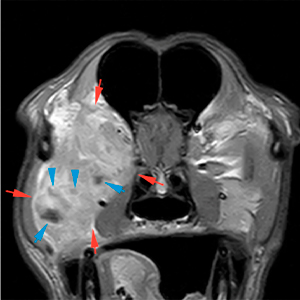
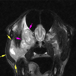
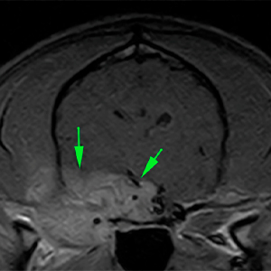
In addition, the mass invades the structures of the orbit, without being able to differentiate the musculature of the ocular globe (pink arrows), extending outwards, invading the masseter muscle (yellow arrows). There is a marked diffuse hyperintensity on T2W of the right temporalis muscle, in the region adjacent to the mass, showing a mild diffuse contrast enhancement (green arrows).
T2W
T2W
T1W+Gd
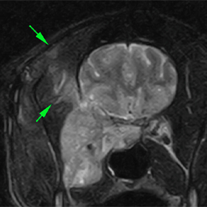
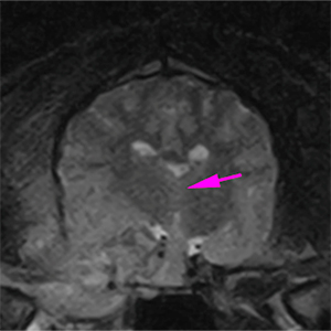
Moreover, the mass causes a marked osteolysis of the rostroventral cranial vault (presphenoid, basisphenoid and temporal), showing a marked intracranial invasion in the region of the right piriform lobe, extending along the base of the calvarium up to the level of the hypophyseal fossa (green arrows). This causes a moderate mass-effect, with a midline shift towards the left (pink arrow). In addition, there is displacement towards the left of other adjacent structures, such as the optic chiasm (orange arrow) and the hypophysis (blue arrow). However, there are no signs of transtentorial or cerebellar herniation.
T2W
T1W+Gd
T2W
T2W
T1W
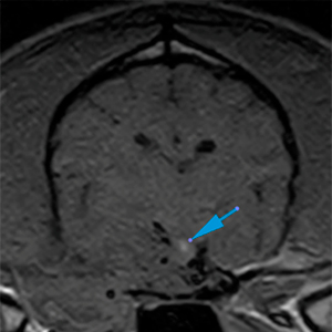
The optic nerve is only visible at its intracranial portion, showing a normal appearance (red arrow). The rest of the optic nerve is embedded within the mass and it is not visible.
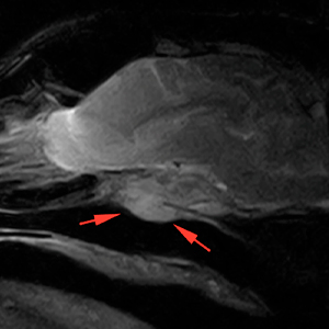
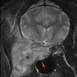
The mass protrudes slightly into the right dorsolateral aspect of the nasopharynx (red arrows).
DIAGNOSIS
Large aggressive mass affecting the right orbit, with intracranial invasion, and extension towards the right masticatory musculature. These findings are most likely consistent with a neoplastic process originating from the orbit (differentials include: rhabdomyosarcoma or myxosarcoma; adenocarcinoma, orbital meningioma, nerve sheath tumour, lymphoma, amongst others).
The intracranial extension of the mass causes a moderated mass-effect, with midline shift and deviation of other adjacent structures, without signs of transtentorial o cerebellar. herniation.
Associated right exophthalmos.
Possible myositis of the temporal muscle vs neoplastic infiltration.
Mild invasion of the nasopharynx.
COMMENTS
Histopathological analysis (biopsy of the outer portion of the mass involving the masseter muscle) is recommended in order to reach a definitive diagnosis, as well as imaging study of the regional lymph nodes and the thoracic cavity for staging.
(Mon. to Fri. 9 a.m. to 6 p.m. gmt+1) Welcome, How can we help you?
We use cookies to ensure that we give you the best experience on our website. If you continue to use this site we will assume that you are happy with it.Ok

No comment yet, add your voice below!