10-year-old female crossbreed. The patient presents a large abdominal mass of unknown origin. On ultrasound the left kidney is not visible.
An abdominal CT scan was performed.
Description
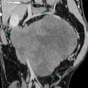
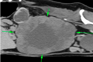
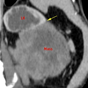
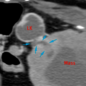
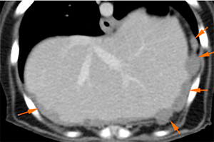
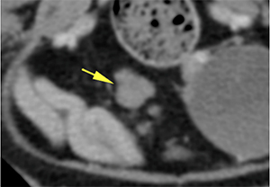
There is a large mass with irregular margins in the left mid-abdomen (green arrows). This mass involves the caudal pole of the left kidney, which is partially distorted (yellow arrow) and ureter. The mass shows heterogeneous soft-tissue attenuation, with a moderate and markedly heterogeneous post-contrast enhancement.
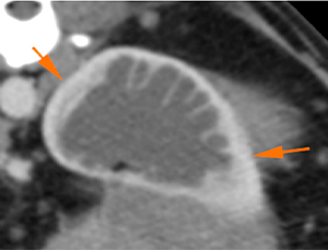
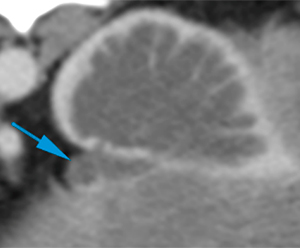
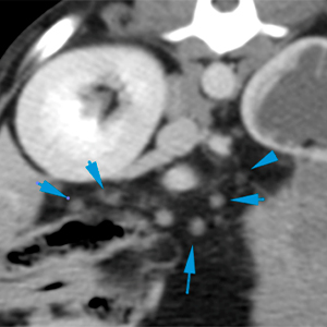
The left kidney shows marked hydronephrosis, presenting only part of the renal cortex and septa, filled with a fluid attenuating material (orange arrows). The proximal portion of the left ureter is also slightly distended (blue arrow), inserting into the mass and embedded within it.
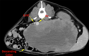
The mass, due to its size, causes a marked mass effect displacing the descending colon and intestinal loops ventrally and to the right, the aorta and caudal vena cava to the right, and the left kidney cranially (yellow arrows).
The caudal vena cava, at the level of the mid-abdomen, is moderately compressed without signs of invasion (green arrows).
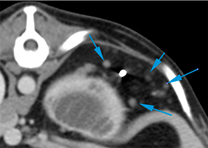
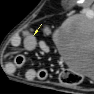
At the level of the peritoneum, there are multiple, variable in size, round nodules, some at the edge of the hepatic parenchyma, being difficult to determine if some of them originate from the liver (orange arrows). However, others are clearly independent, distributed throughout the peritoneal cavity (blue arrows).
Multiple lymph nodes (mesenteric, splenic) are enlarged, with heterogeneous morphology and marked contrast enhancement (yellow arrows).
Diagnosis
Large mass in the left mid-abdomen, involving the left kidney/ureter, consistent with an aggressive neoplastic process (carcinoma, sarcoma, among others), most likely. This mass produces hydronephrosis and mild hydroureter, due to invasion/obliteration of the ureter.
The mass produces a marked mass-effect on multiple structures, including compression of the caudal vena cava, without evident invasion of the latest.
Multiple peritoneal and retroperitoneal nodules, consistent with neoplastic infiltration (carcinomatosis/sarcomatosis).
Moderate lymphadenopathy of multiple abdominal lymph nodes, consistent with metastasis most likely.
Comments
FNAs/histopathological analysis of the mass is recommended to reach a definitive diagnosis. However, given the findings in the abdominal cavity, metastasis is quite likely. Aspiration of some of the peritoneal nodules and/or lymph nodes is also recommended to try to reach a definitive diagnosis. Consider imaging study of the thoracic cavity for staging.
(Mon. to Fri. 9 a.m. to 6 p.m. gmt+1) Welcome, How can we help you?
We use cookies to ensure that we give you the best experience on our website. If you continue to use this site we will assume that you are happy with it.Ok


No comment yet, add your voice below!