13-year-old female neutered crossbreed. History of tremors and weakness in the hindlimbs, aggravated over the last 8 days.
A head CT scan was performed.
Description
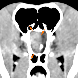
In the right rostral cranial fossa, at level of the right olfactory bulb and frontal lobe, there is a round, well-defined lesion, showing a marked contrast enhancement (orange arrows). This lesion has an extra-axial location, with a broad base in the ethmoidal fossae, causing a marked midline shift towards the left.
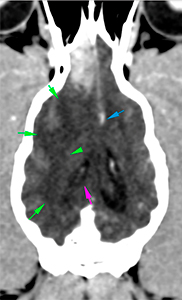
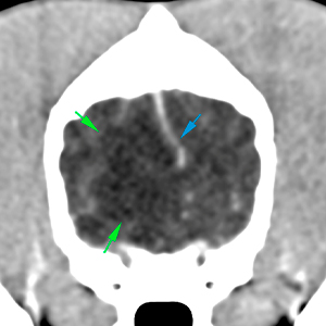
Caudal to the lesion, there is an ill-defined, hypoattenuating, non-contrast enhancing, lesion extending along the white matter throughout the right hemisphere, up to the most rostral portion of the right occipital lobe, which could be consistent with an extensive area of perilesional vasogenic oedema (green arrows). This lesion causes a mass-effect with midline shift towards the left (blue arrow), and compression of the right lateral ventricle (pink arrow), which is markedly reduced in size in comparison to the contralateral side. There is no evidence of cerebellar or transtentorial herniation.
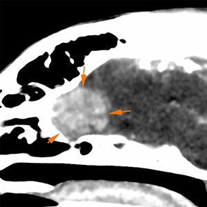
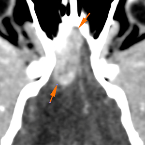
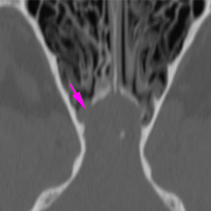
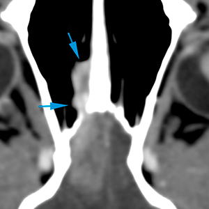
The cribriform plate, adjacent to the previously described mass, shows a mild lysis (pink arrow). In addition, there is a small soft tissue attenuating and strong contrast-enhancing lesion adjacent to the right cribriform plate and extending slightly rostrally, in contact with the nasal septum (blue arrows).
Diagnosis
Single extra-axial intracranial mass, in the right olfactory bulb/ frontal lobe, with possible involvement of the cribriform plate and extension into the right nasal cavity. The changes are most likely consistent with a neoplastic process (esthesioneuroblastoma/olfactory neuroblastoma, most likely; others like meningioma, histiocytic sarcoma, oligodendroglioma are less likely). Other differentials, such as abscess or granuloma, are very unlikely.
Extensive perilesional vasogenic oedema, extending to the right occipital lobe, causing mass-effect and compression of the right lateral ventricle.
Comments
Histopathological analysis of the mass is necessary in order to achieve a definitive diagnosis.
(Mon. to Fri. 9 a.m. to 6 p.m. gmt+1) Welcome, How can we help you?
We use cookies to ensure that we give you the best experience on our website. If you continue to use this site we will assume that you are happy with it.Ok


No comment yet, add your voice below!