Mass in right tympanic bulla with nasopharyngeal, periaural soft tissues and intracranial invasion in a cat
1-year-old, male, Blue Russian cat was presented with nasal and otic discharge. The patient has non-regenerative anaemia and is positive for feline leukaemia virus (FeLV). A CT of the head was performed.
Description
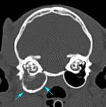
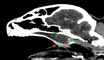
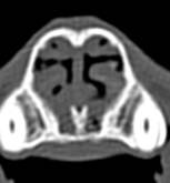
There is asymmetry of the right tympanic bulla with mild expansion and irregular thickening of its wall (blue arrows). There is hypoattenuating material (57HU) inside its lumen, affecting both compartments, with slight erosion of the septum.
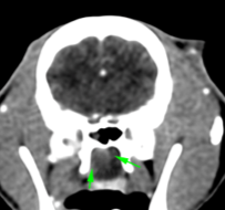
There is an ill-defined lesion extending from the auditory tube (Eustachian tube), which is widened, towards the nasopharynx, where it forms a mass (green arrows). This lesion is hypoattenuating (30HU) and shows a mild post-contrast enhancement (45HU), mainly peripheral. This results in complete obstruction of the nasopharynx and displacement of the soft palate ventrally (red arrows). The lesion is lateralised to the right, invading the soft tissues located medial to the temporomandibular joint (pink arrow) and rostral to the bulla. There is a slight accumulation of hypoattenuating material in the horizontal part of the right external ear canal. No alterations are seen in the left ear.
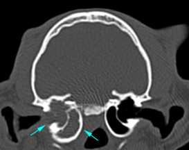
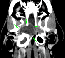
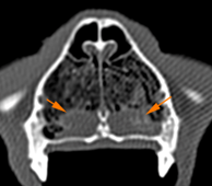
There is an enlargement of the right foramina at the base of the skull (yellow arrows) (oval and rostral alar foramina; and orbital fissure). They are in contact with the nasopharyngeal mass suggesting a possible invasion of these, which contain material isoattenuating to the mass (62HU). In addition, there is a discrete asymmetry, with ill-defined margins, at the level of the right trigeminal ganglion, without evidence of compression of the adjacent parenchyma (orange arrows).
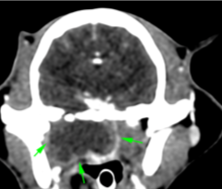
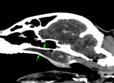
There is a moderate amount of hypoattenuating material (44HU) in the nasopharynx (green arrows), rostral to the previously described mass (m). There is also hypoattenuating material (25HU) in the ventral aspect of both nasal cavities (orange arrows) and choanae, with moderate mucosal enhancement.
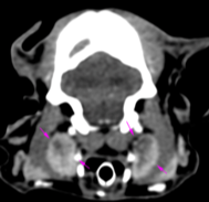
Both mandibular and medial retropharyngeal lymph nodes are prominent with heterogeneous contrast enhancement (40-60HU), with hypoattenuating areas (pink arrows).
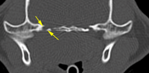
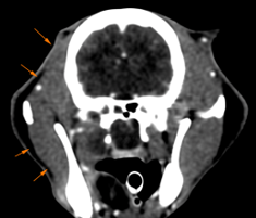
Secondary to the previously described lesion, there is mild atrophy of the right masticatory muscles (temporal and masseter right muscles -orange arrows-).
Diagnosis
Mass in the right tympanic bulla with invasion of the nasopharynx and periaural soft tissues, with possible involvement of the trigeminal nerve and intracranial invasion. These findings are most likely consistent with a neoplastic process (e.g., lymphoma being FELV+). Other differentials, such as an inflammatory/infectious or granulomatous process cannot be ruled out, although less likely. Secondary to this lesion, there is:
Accumulation of material in the rostral nasal cavity and nasopharynx.
Right masticatory muscle atrophy, possibly due to ipsilateral trigeminal nerve involvement/damage.
Bilateral mandibular and medial retropharyngeal lymphadenopathy, metastasis most likely vs reactive.
Comments
Considering that the patient is FELV+, it is likely that the lesions described are of neoplastic origin (lymphoma). Sampling of the mass is required to reach a definitive diagnosis. Magnetic resonance imaging is recommended for a better evaluation of the nervous system.
(Mon. to Fri. 9 a.m. to 6 p.m. gmt+1) Welcome, How can we help you?
We use cookies to ensure that we give you the best experience on our website. If you continue to use this site we will assume that you are happy with it.Ok


No comment yet, add your voice below!