Metaphyseal osteopathy (hypertrophic osteodystrophy) in a puppy
5-months-old female Weimaraner with fever, vomiting, cough and joint pain, as well as pain when opening the mouth.
A full body CT-scan was performed.
Description
Thoracic limbs
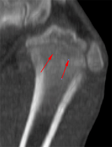
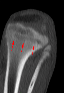
There are multiple moth-eaten areas of bone lysis creating a band running parallel to the physis, surrounded by a hyperattenuating ill-defined region (yellow arrows) in the proximal metaphysis of both humerus (right>left).
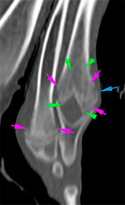
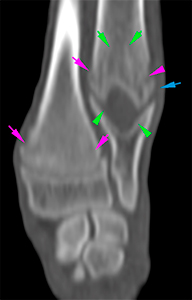
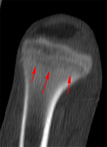
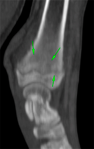
The distal metaphysis of both radius and ulna shows the same lesions (right>left). There are multiple moth-eaten areas of bone lysis creating a band running parallel to the physis surrounded by a hyperattenuating region with ill-defined margins (pink arrows) . There is a mild solid and irregular periosteal reaction at the distal metaphysis of the ulna (blue arrow) and radius (red arrows).
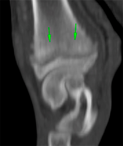
In both forelimbs, there is a hypoattenuating candle flame-like area extending from the distal physis of the ulna, proximally along the metaphysis and distal diaphysis (green arrows).
Left Forelimb
Right Forelimb
There are no signs of joint effusion, joint incongruity or periarticular soft tissue swelling.
Pelvic limbs
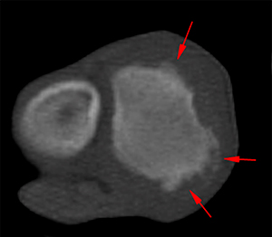
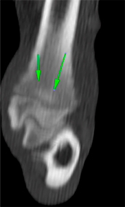
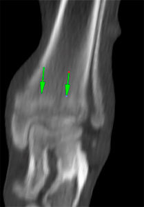
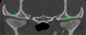
There are similar lesions as the ones described in the thoracic limbs, adjacent to all physis, more severe at the proximal (red arrows) and distal metaphysis (green arrows) both tibiae (R>L).
Left hindlimb
Right hindlimb
There are no signs of joint effusion, joint incongruity or periarticular soft tissue swelling.
Head
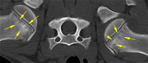
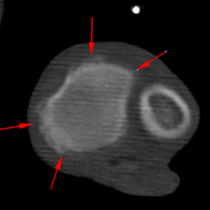
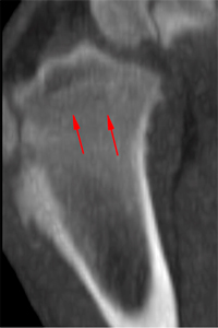
There are multiple moth-eaten areas of lysis in both mandibular condyles and a mild irregularity of the articular surface (green arrows).
Diagnosis
Areas of moth-eaten bone lytic lesions and sclerotic area running parallel to the physis, in the metaphyseal regions of the humerus, ulna, radius, femur and tibia, more evident on the right limb; and lytic lesions in both mandibular condyles. These lesions are most likely consistent with metaphyseal osteopathy (hypertrophic osteodystrophy). Other differential diagnosis, such as incidental anatomical variation associated with rapid growth and remodeling of the metaphyseal regions, considering the age and breed of the patient, or an infectious process (metaphyseal osteomyelitis) cannot be completely ruled out, although less likely.
Findings in the distal metaphyseal regions of both ulnae, consistent with bilateral retained cartilaginous core.
Comments
The findings described in the appendicular skeleton, together with the patient’s age, clinical signs and breed, are highly suggestive of metaphyseal osteopathy (hypertrophic osteodystrophy). However, an incidental anatomical variation associated with rapid growth, considering the patient’s age and breed, cannot be completely ruled out, although less likely.
No abnormalities are seen in the joints, although, arthrocentesis could be considered for sampling, including culture, to reach a definitive diagnosis given the joint pain described in the clinical history.
The following articles might be of interest:
Hill M, Scudder C, Glanemann B, Drees R. Hypertrophic osteodystrophy in a dog imaged with CT. Rec. Case Rep. 2015; 3(1). doi: 10.1136/vetreccr-2014-000155.
(Mon. to Fri. 9 a.m. to 6 p.m. gmt+1) Welcome, How can we help you?
We use cookies to ensure that we give you the best experience on our website. If you continue to use this site we will assume that you are happy with it.Ok


No comment yet, add your voice below!