Osseous defect in the caudal portion of the hard palate
6-year-old male neutered cat. History of chronic sneezing produced by a palatal defect caused by previous trauma. Despite surgical interventions the symptoms persist.
A head CT scan was performed.
Description
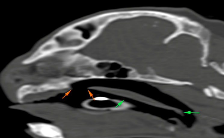
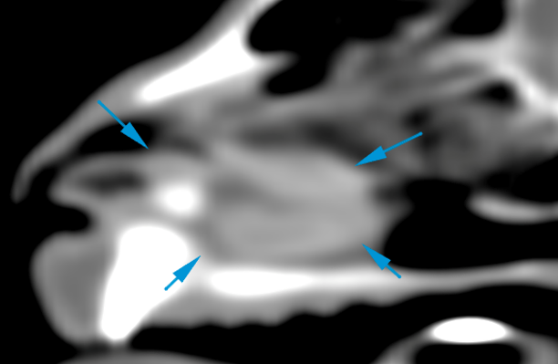
There is a bone defect, with fusiform shape, in the caudal and medial aspect of the hard palate, at the level of the horizontal plate of the palatine bone (orange arrows). This defect extends from the molar region towards the most caudal aspect of the palatine bone, where the soft palate begins, remaining intact (green arrows). In addition, this defect communicates the oral cavity with the rostral portion of the nasopharynx and both choanae.
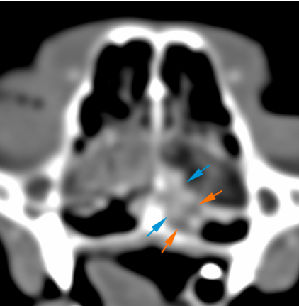
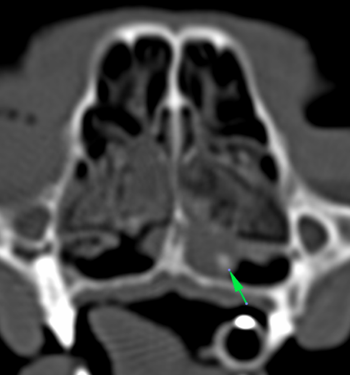
The left choana is partially obliterated by a fluid/soft tissue attenuating material (orange arrows), and multiple small mineral foci (green arrow). After contrast administration, the nasal mucosa, in this region, is mildly thickened, showing a marked contrast enhancement (blue arrows).
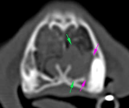
There is a focal area of soft tissue attenuation (green arrows), extending along the ventral aspect of the left nasal cavity, up to the level of the left upper canine. This is associated with focal lysis of the nasal turbinates. Rostrally, there is a round, mineral attenuating structure, adjacent to the root of the left upper canine (red arrows). Associated with this lesion, there is a mild lysis of the maxillary cortex that surrounds the periapical region of the left canine (pink arrows).
After contrast administration, this region shows a marked and homogeneous contrast enhancement, with a marked and focal thickening of the nasal mucosa (blue arrows) (red arrows- mineral attenuating structure mentioned above).
Diagnosis
Osseous defect in the caudal portion of the hard palate, probably secondary to an old trauma described in history. This defect causes an abnormal communication of the oral cavity with the rostral portion of the nasopharynx and choanae.
Mineral attenuating structure located in the rostroventral aspect of the left nasal cavity and multiple mineral attenuating foci along the left choana, which could be consistent with foreign bodies most likely or rhinoliths associated with chronic rhinitis.
Findings in the left nasal cavity associated with the previously described structures are consistent with an inflammatory/infectious process (rhinitis associated with foreign body vs other types of non-specific rhinitis). A neoplastic process is unlikely, although it cannot be completely ruled out.
Comments
The findings in the left nasal cavity are consistent with presence of foreign bodies and associated chronic rhinitis, most likely related to the defect of the hard palate, and consequent abnormal communication of the oral cavity with the nasopharynx and choanae. Rhinoscopy and sampling of the lesion at the left nasal cavity is required to reach a definitive diagnosis.
(Mon. to Fri. 9 a.m. to 6 p.m. gmt+1) Welcome, How can we help you?
We use cookies to ensure that we give you the best experience on our website. If you continue to use this site we will assume that you are happy with it.Ok


No comment yet, add your voice below!