6-months-old male spanish hound. Three weeks ago, he presented with tachypnoea, fever, and apathy; he was diagnosed with bronchopneumonia. A blood culture was performed and Pasteurella canis was isolated. A few days ago, he started with painful joints and thoracolumbar spine.
A CT scan of fore limbs, pelvic limbs, thoracolumbar spine and thorax was performed.
Description
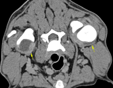
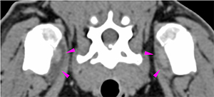
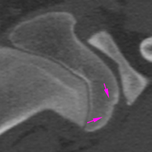
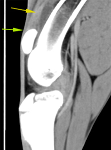
There is joint effusion in both shoulders (yellow arrows) that extends distally along the bicipital tendon sheath (pink arrowheads), more prominent in the right shoulder. There is moderate thickening of the periarticular soft tissues, showing a diffuse contrast enhancement.
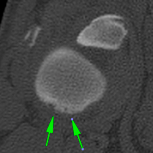
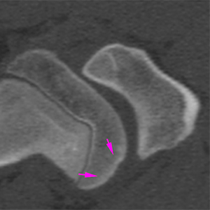
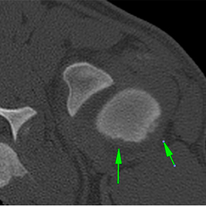
In both humeral heads, there are multiple concave well-defined subchondral bone defects at the caudal aspect of the humeral head (green arrows) surrounded by an area of diffuse sclerosis (pink arrows). These changes are more prominent in the left shoulder.
Right shoulder
Left shoulder
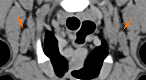
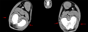
The axillary lymph nodes are slightly prominent showing homogeneous contrast enhancement (orange arrows).
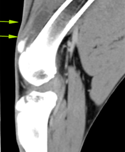
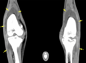
Marked joint effusion in both stifles (R>L) (yellow arrows), extending from the distal femoral diaphyseal portion along the tendon sheath of the long digital extensor muscle. The periarticular soft tissues are moderately thickened, showing homogeneous post-contrast enhancement (red arrows).
Right stifle
Left stifle
There are no evident abnormalities at the spine included.
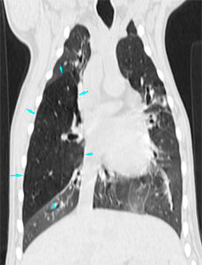
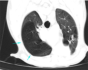
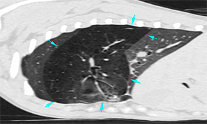
The pulmonary parenchyma is well expanded. The right middle lung lobe (blue arrows) is severely enlarged (hyperinflated), with decreased homogeneous attenuation (-1000 HU) compared to the rest of the lung lobes (-800 HU), causing displacement of the adjacent lung lobes and mild mediastinal shift towards the left.
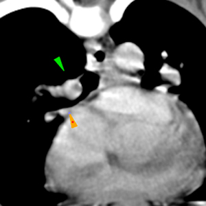
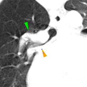
The lobar bronchi of the right middle lung lobe is focally narrowed (orange arrowhead), with a moderate obliteration of its lumen at its most proximal aspect, adjacent to the subjectively prominent right lobar pulmonary artery (green arrows). This artery, however, is similar in size to the left one and has a homogeneous contrast enhancement, with no filling defects. The bronchi can be followed to the periphery, filled with air, and no changes in the bronchial walls are seen.
Diagnosis
Thickening of the periarticular soft tissues of the shoulders and stifles, with associated joint effusion. These findings are consistent with an inflammatory/infectious process (septic or immune-mediated polyarthritis, most likely); although drug-induced polyarthritis cannot be ruled out considering the history of the patient.
Subchondral bone defects in both humeral heads (L>R), can be consistent with erosive arthritis vs. osteochondrosis could be also possible given the location.
Mild axillary lymphadenopathy, most likely reactive.
Hyperinflation and decreased attenuation of the right middle lung lobe, consistent with lobar emphysema, most likely congenital in origin given the age of the patient; although acquired after the bronchopneumonia described in the history, it cannot be completely ruled out.
Comments
Follow up: Treatment with doxycycline was started prior to sampling. Cerebrospinal fluid analysis was normal. Arthrocentesis of the shoulders and stifles was performed, showing neutrophils with no apparent bacteria. The culture was negative. The patient improved with antibiotic therapy and analgesic treatment. Fifteen days later, the physical examination was completely normal.
(Mon. to Fri. 9 a.m. to 6 p.m. gmt+1) Welcome, How can we help you?
We use cookies to ensure that we give you the best experience on our website. If you continue to use this site we will assume that you are happy with it.Ok


No comment yet, add your voice below!