9-year-old male Doberman, with a mass in the left tarsal area, that affects the bone with osteolysis. For months he has been intermittently lame of the left pelvic limb. The mass has increased in size.
A CT of the thorax, abdomen and hind limbs was performed.
Description
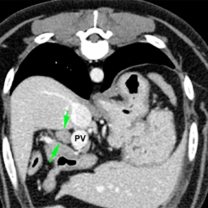
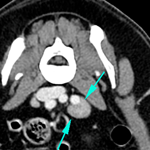
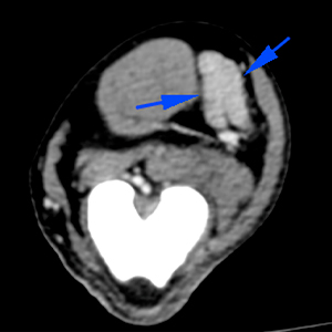
The hepatic lymph nodes are enlarged with homogeneous contrast enhancement (green arrows). The left medial iliac lymph node is also enlarged (blue arrows).
Moderate atrophy of the musculature of the left pelvic limb (red arrows).
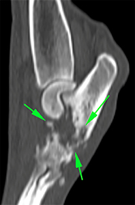
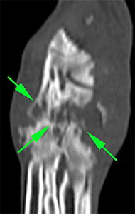
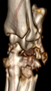
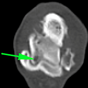
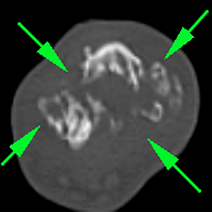
Aggressive, polyostotic and mainly osteolytic bone lesion affecting the left tarsi, including the distal aspect of the calcaneus and talus, and proximal aspects of the central tarsal and 4th tarsal bones, mainly affecting the articular surfaces of the proximal intertarsal joint, with irregular lysis and ill-defined margins of the subchondral bone (green arrows). There is a moderate periosteal reaction around the calcaneal tuberosity. There is a hypoattenuating lesion at the medial and proximal aspect of the subchondral bone of the talus.
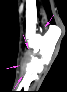
The osseous changes are associated with a hypoattenuating soft tissue mass (50 HU), which shows a marked contrast enhancement (120HU) (pink arrows). The tarsocrural and tarso-metatarsal joints are intact.
Moderate enlargement of the left popliteal lymph node (blue arrows).
Diagnosis
Polyostotic aggressive lesion in the left tarsus with involvement of the proximal intertarsal joint. This lesion is most likely consistent with neoplasia (histiocytic sarcoma or synovial sarcoma). Others, such as infectious process (septic arthritis) or a very severe inflammatory process, are less likely.
Ipsilateral popliteal, medial iliac and hepatic lymphadenopathy, consistent with metastasis vs reactive.
Comments
Cytology or histopathological analysis of the tarsus and FNAs of the affected lymph nodes are recommended in order to reach a definitive diagnosis.
(Mon. to Fri. 9 a.m. to 6 p.m. gmt+1) Welcome, How can we help you?
We use cookies to ensure that we give you the best experience on our website. If you continue to use this site we will assume that you are happy with it.Ok


No comment yet, add your voice below!