14-years-old female Siberian Husky. An acute fracture of the right femur was seen. A CT of the right femur and thorax was performed. A thoracolumbar spine and thoracic CT scan was performed.

Description
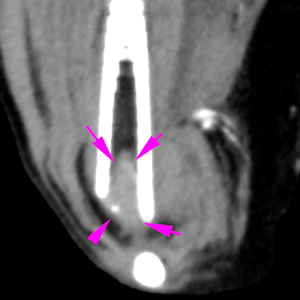
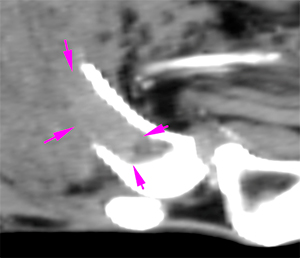
There is a complete and oblique fracture of the distal diaphysis of the right femur, with caudomedial displacement of the distal aspect of the right femur (blue arrows). There is marked permeative osteolysis along the distal diaphysis of the femur, with disruption and thinning of the cortex (green arrows) with a wide transition zone. At this level, there is a soft tissue lesion with moderate contrast enhancement affecting the medullary cavity of the bone (pink arrows).
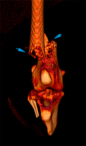
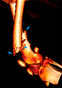
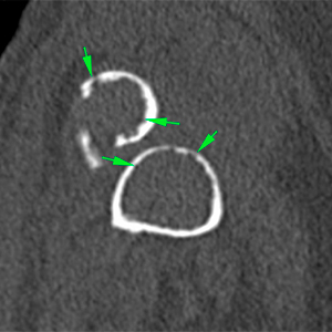
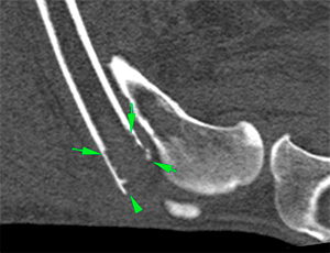
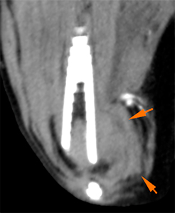
There is a thickening of the soft tissues adjacent to the fracture with non-contrast enhancement, most marked in the medial aspect of the femur (orange arrows).
In the thorax, there are multiple, variable in size, well-defined, soft tissue nodules distributed throughout the pulmonary parenchyma. The biggest nodules (2 cm approximately) are located in the right caudal and accessory lung lobes (orange arrows).
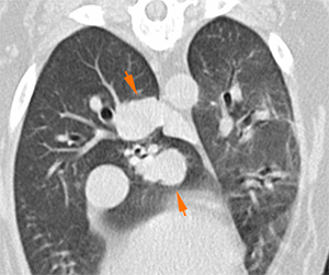
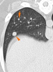
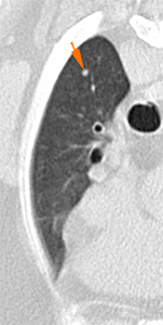
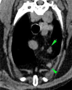
The cranial mediastinal and sternal lymph nodes are mildly enlarged (green arrows).
Diagnosis
- Aggressive bone lesion in the distal diaphysis of the right femur with associated pathological fracture: most likely consistent with neoplasia (primary bone tumor –osteosarcoma, most likely).
- Thickening of the soft tissues at the medial aspect of the right femur, most likely consistent with inflammation related to the fracture.
- Multiple pulmonary nodules, most likely consistent with metastasis.
- Mild sternal and cranial mediastinal lymphadenopathy: reactive/metastasis.

No comment yet, add your voice below!