2-year-old, male neutered, American Staffordshire terrier.
Previous history of haematuria. Castrated a few months ago on suspicion of prostatic problems. For the past 2 weeks, marked weight loss, apathy, vomiting and anorexia.
A CT scan was performed.
Description
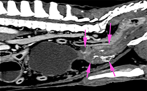
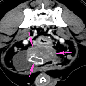
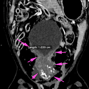
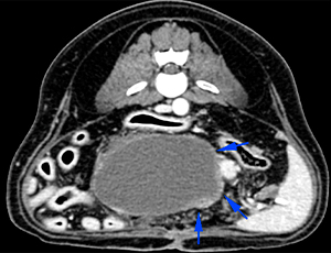
There is a mass with heterogeneous attenuation and ill-defined margins, located caudal to the urinary bladder, at the level of the prostate, with slight cranial extension towards the trigone of the urinary bladder. The latest shows a thickened and irregular surface (blue arrows). The mass is hypoattenuating, with multiple dystrophic mineralizations, and has a marked and heterogeneous contrast enhancement (pink arrows).
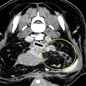
Moderate perilesional inflammation of the adjacent fat with small peritoneal nodules, showing a heterogeneous contrast enhancement (yellow circle).
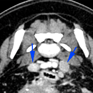
The medial iliac lymph nodes are enlarged, with slightly heterogeneous contrast enhancement (blue arrows).
Diagnosis
Prostatic lesion with possible invasion of the trigone of the urinary bladder, consistent with a neoplastic process (e.g. prostatic carcinoma, adenocarcinoma, etc.), most likely. Other causes such as severe chronic prostatitis (infectious or inflammatory) are less likely.
The adjacent peritoneal changes are suggestive of tumoral infiltration (carcinomatosis) or focal peritonitis.
Medial iliac lymphadenopathy, metastasis vs reactive.
(Mon. to Fri. 9 a.m. to 6 p.m. gmt+1) Welcome, How can we help you?
We use cookies to ensure that we give you the best experience on our website. If you continue to use this site we will assume that you are happy with it.Ok

No comment yet, add your voice below!