Psoas muscle abscess associated with a foreign body
2-year-old male American Staffordshire Terrier with history of lumbosacral pain.
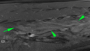
A lumbosacral MRI was performed.
DESCRIPTION
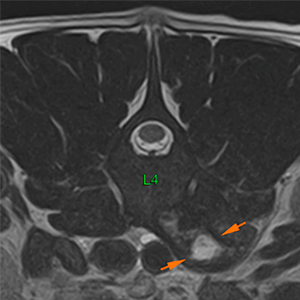
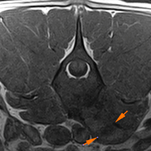
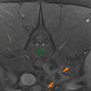
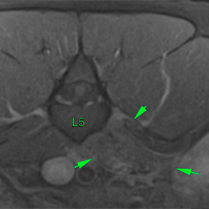
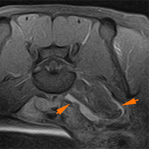
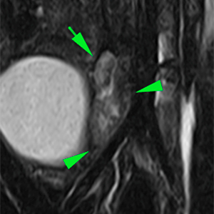
At the level of the left psoas minor muscle, there is a lesion extending from the cranial aspect of L3 to the caudal aspect of L5 (orange arrows). The muscle is markedly enlarged, presenting an oval-shaped lesion with irregular and well-defined margins. The lesion has a hyperintense center on T2W and hypointense on T1W, showing a marked peripheral enhancement with persistence of a central hypointense area, consistent with a cavitary lesion.
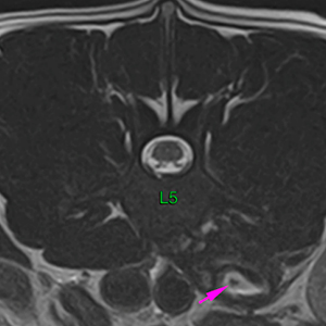
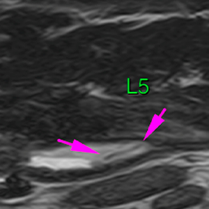
In the caudal aspect of the lesion, there is a linear hypointense structure (pink arrow).
T2W
T1W
T1WFS+Gd
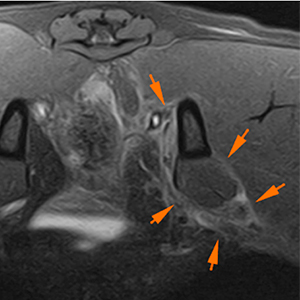
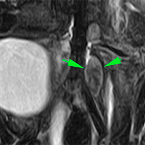
There is a diffuse hyperintensity on T2W, with marked contrast enhancement, of the left minor and major psoas muscles surrounding the lesion previously described, which extends from the ventral aspect of L2 to the most caudal aspect of the musculature, at the level of the iliopsoas muscle (green arrows).
More caudally, there is a marked enhancement of the fascial tissues that surround the left iliopsoas muscle and extend along the femoral area, as well as of the left intrapelvic area (orange arrows).
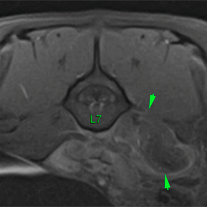
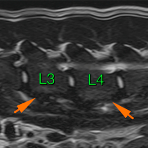
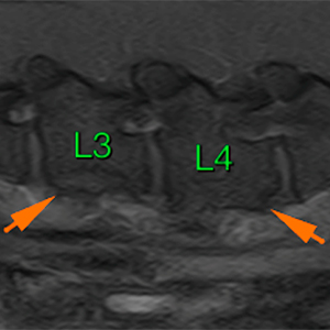
The vertebral bodies of L3 and L4, at their ventral aspect, show a marked solid periosteal reaction, with slightly irregular margins, being more severe at the level of L3 (orange arrows).
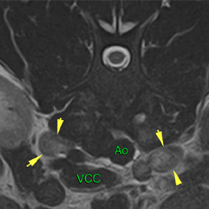
Some of the lumbar aortic and medial iliac (yellow and green arrows respective) lymph nodes are enlarged, showing homogeneous intensity and moderate contrast enhancement.
DIAGNOSIS
Lesion at the level of the left psoas muscle, consistent with an abscess (probably associated with a migratory foreign body, which appears to be located in the caudal aspect of the lesion i.e., grass awn, splinter, etc) most likely. Other differentials, such as a neoplastic process affecting the musculature, are unlikely, although they cannot be completely ruled out.
Marked myositis of the left psoas muscle and oedema/inflammation of the adjacent soft tissues (intrapelvic and femoral tissues on the left and ventromedial aspect). Neoplastic infiltration is unlikely.
Marked periosteal reaction at the ventral aspect of L3 and L4, consistent with inflammatory changes associated with the possible abscess.
Moderate lumbar aortic and medial iliac lymphadenopathy, most likely reactive. Metastasis is unlikely.
COMMENTS
The presence of a grass awn inside a muscular abscess was confirmed by ultrasound. The foreign material was surgically removed.
(Mon. to Fri. 9 a.m. to 6 p.m. gmt+1) Welcome, How can we help you?
We use cookies to ensure that we give you the best experience on our website. If you continue to use this site we will assume that you are happy with it.Ok


No comment yet, add your voice below!