14-year-old male dog, with an adrenal mass.
An abdominal CT scan was performed.
Description
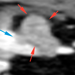
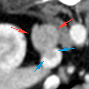
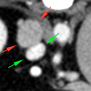
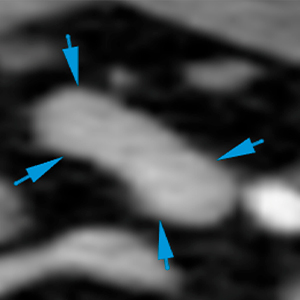
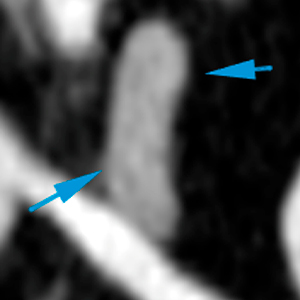
The right adrenal is enlarged, presenting a round mass at its cranial pole with ill-defined margins, showing a moderate and heterogeneous contrast enhancement (red arrows). At its cranial aspect, the mass shows a pedunculated shape that invades the caudal vena cava, occupying approximately 50% of its diameter (blue arrow). In addition, at its medial aspect, the mass is in close contact with the caudal vena cava, slightly compressing it and reducing its diameter (green arrows).
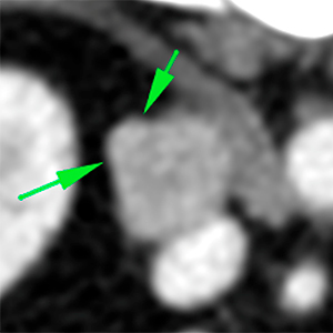
The phrenicoabdominal vein is moderately thickened in its path near the adrenal mass, showing a soft tissue attenuation, similar to the mass, consistent with vascular invasion (green arrows).
The left adrenal has a normal size and homogeneous attenuation (blue arrows).
Diagnosis
Right adrenal mass with vascular invasion of the caudal vena cava and phrenicoabdominal vein, consistent with neoplasia (adenocarcinoma, pheochromocytoma, most likely).
(Mon. to Fri. 9 a.m. to 6 p.m. gmt+1) Welcome, How can we help you?
We use cookies to ensure that we give you the best experience on our website. If you continue to use this site we will assume that you are happy with it.Ok


No comment yet, add your voice below!