13-year-old male neutered crossbreed. Previous history of cough and mild anorexia. X-rays revealed a possible mediastinal mass.
A thoracic CT scan was performed.
Description
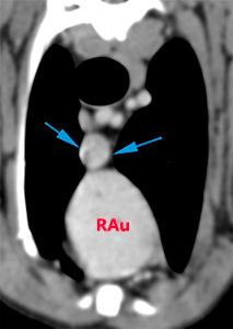
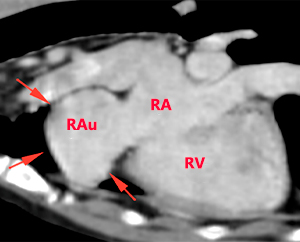
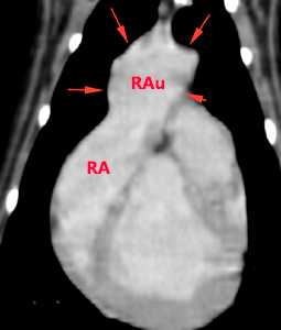
There is moderate/marked dilatation of the right auricle (red arrows), that appears as a structure with a very thin wall, homogeneously filled with contrast, connected to the right atrium, and prolapsing towards the cranioventral mediastinum located ventral to the cranial vena cava (blue arrows). There is no filling defect or other abnormalities within the right auricle.
Diagnosis
Moderate right auricle aneurysm, with mild prolapse towards the cranioventral mediastinum. The aetiology of this finding is uncertain, although it is described that this could be associated with a focal defect in the pericardium and concurrent herniation of the right auricle, or it could also be unrelated to a pericardial defect (secondary to inflammatory alterations, among others). The clinical significance of this finding is uncertain.
Comments
Cardiac evaluation, including ECG and echocardiography, is recommended to try to detect other alterations associated to the findings in the right auricle.
(Mon. to Fri. 9 a.m. to 6 p.m. gmt+1) Welcome, How can we help you?
We use cookies to ensure that we give you the best experience on our website. If you continue to use this site we will assume that you are happy with it.Ok


No comment yet, add your voice below!