12-year-old male Maltese with anorexia. During the physical examination, a submandibular mass was found. The ultrasound revealed anechoic content inside the mass, and the cytology showed a high neutrophil count.
A neck and thoracic CT scan was performed.
Description
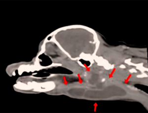
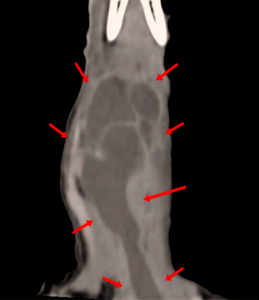
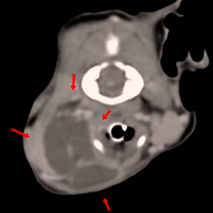
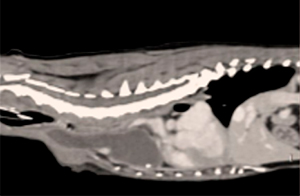
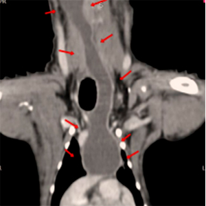
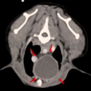
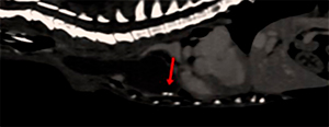
There is a cavitary lesion, with fluid, non-enhancing content, with encapsulated appearance with well-defined and smooth margins, showing a mild peripheral contrast enhancement. This lesion extends from the most caudal aspect of the base of the tongue through the submandibular, retropharyngeal and cervical areas, caudally up to the cranial and ventral aspect of the thorax, at the level of the cranial mediastinum.
At the level of the mandibular salivary glands, there is vascularized tissue, showing contrast enhancement, in the dorsal aspect of the cystic lesion, in the right retropharyngeal area: this tissue shows an enhancement similar to the parenchyma of the salivary glands. The cystic lesion displaces the right mandibular salivary gland laterally.
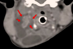
There are two small mineralized foci at the ventral aspect of the cranial mediastinal portion of the cystic lesion. These could be mineral foci with a dystrophic origin, but calculi cannot be completely ruled out (sialoliths that have migrated caudally).
Diagnosis
Cystic structure in the submandibular, cervical and thoracic area consistent with a sialocele, originating from a portion of the right mandibular salivary gland, or a branchial cyst. A neoplasitc process is unlikely, given the characteristics of the lesion.
(Mon. to Fri. 9 a.m. to 6 p.m. gmt+1) Welcome, How can we help you?
We use cookies to ensure that we give you the best experience on our website. If you continue to use this site we will assume that you are happy with it.Ok


No comment yet, add your voice below!