5-year-old, male neutered. American Staffordshire Terrier, came to the emergency room after being run over by a train, with an open wound on the left cheek with exposure of the masseter muscles.
A CT scan of the head was performed to assess the extent of the injuries.
Description
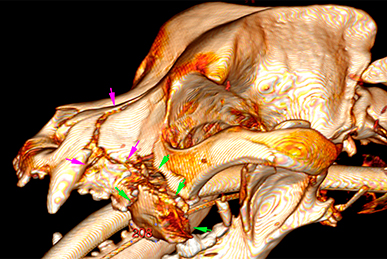
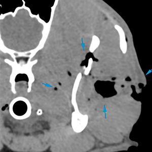
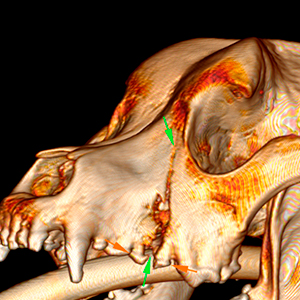
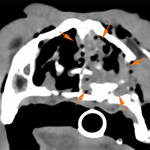
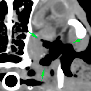
In the left facial area, there is an extensive lesion with disruption of the skin, with a large defect, that extends from the maxillary region up to the temporomandibular joint (red arrows). Furthermore, in this area, there is thickening of all the subcutaneous tissues, with presence of gas located between the masticatory muscles (masseter, temporal and pterygoid) (blue arrows).
Associated with this injury, there are multiple fractures:
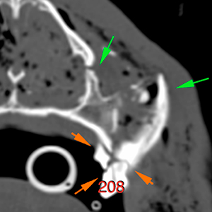
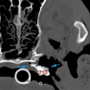
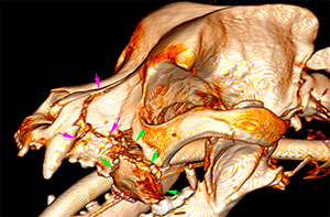
Comminuted fracture of the left maxilla, with multiple fracture lines and bone fragments with ventrolateral displacement (pink, green and blue arrows). The left upper 4th premolar (208) is fractured (orange arrows).
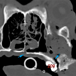
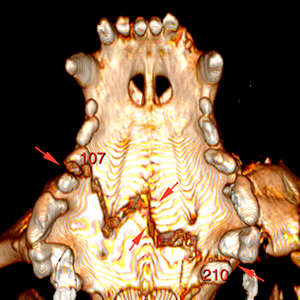
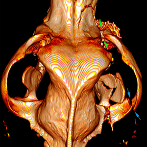
Fracture line at the level of the right maxilla (green arrows). Fracture of the right upper 3rd premolar (107) (orange arrows).
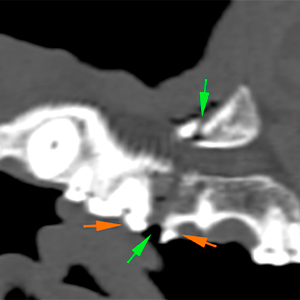
The fracture lines that affect the palatine process of both maxillae converge in a zigzag fracture line along the osseous palate (red arrows).
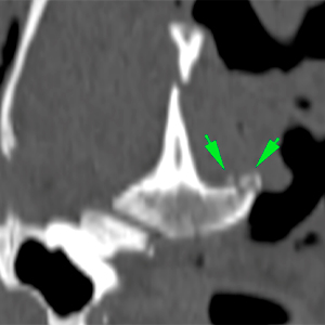
There is a comminuted fracture of the left ramus of the mandible, affecting the masseteric fossa and coronoid process (blue arrows).
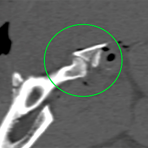
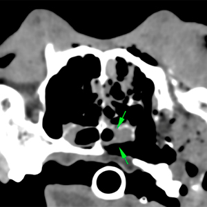
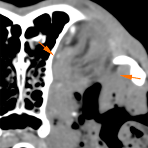
There are multiple fractures at the level of the left temporomandibular joint (blue and green arrows). However, the temporomandibular joint appears to be congruent (green circle).
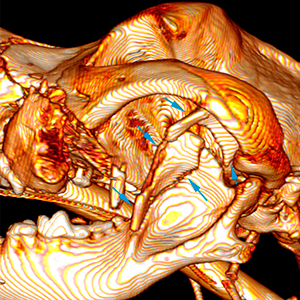
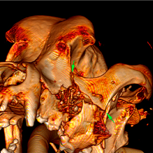
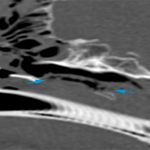
There are two complete fractures affecting the left zygomatic arch. One at its rostral aspect, at the level of the zygomaticomaxillary suture (green arrows), and another at its caudal aspect, at the level of the zygomatic process of the temporal bone (blue arrows).
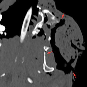
Associated with these fractures, there is a moderate amount of hypoattenuating material in both nasal cavities (L>R) (orange arrows). There is a slight narrowing of the left choana (green arrows), with presence of a mild amount fluid.
There is a mild amount of gas along the left side of the soft palate, in its dorsal aspect (blue arrows), consistent with a laceration, without clear communication between the nasopharynx and the oral cavity.
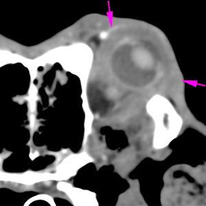
At the level of the left orbit, there is a large amount of gas in the retrobulbar space (green arrows), as well as a mild thickening of the soft tissue structures of the retrobulbar space, with a mild loss of visualization of the retrobulbar fat (orange arrows). Rostral exophthalmos, with thickening and increased attenuation of the periocular fat, while the eyeball remains intact (pink arrows).
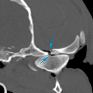
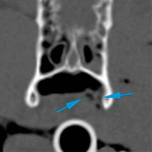
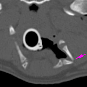
Complete fracture of the left epihyoid bone (pink arrow).
conclusions
Tear/open wound in the left maxillary/masseteric region, with marked subcutaneous emphysema along the facial region.
Multiple fractures affecting:
Right maxilla, with fracture of 107;
Left maxilla, with displacement of multiple bone fragments and fracture of 208;
Fracture of the osseous palate and possible tear of the soft palate, without evident communication of the nasal cavity or nasopharynx with the oral cavity;
Fracture of the left mandibular ramus;
Fracture of the left temporomandibular joint, without obvious signs of incongruity;
Fracture of the left zygomatic arch;
Fracture of the left epihyoid bone.
Nasal and left choana secretion, compatible with haemorrhage.
Emphysema and inflammation at the retrobulbar space, with left exophthalmos.
(Mon. to Fri. 9 a.m. to 6 p.m. gmt+1) Welcome, How can we help you?
We use cookies to ensure that we give you the best experience on our website. If you continue to use this site we will assume that you are happy with it.Ok

No comment yet, add your voice below!