5-years-old female neutered Dogue de Bordeaux. The dog has a history of chronic digestive symptoms and weight loss. Recently the symptoms worsened. Because of the progression of the symptoms, an abdominal ultrasound was performed, revealing a splenic mass which, on cytological examination, is suspected to be histiocytic sarcoma. Due to the findings, an abdominal CT scan was performed.
Description
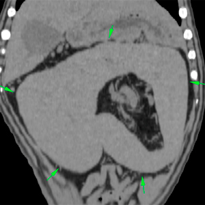
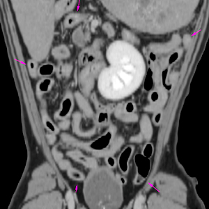
The splenic parenchyma is markedly enlarged, with moderately rounded margins and an abnormal position, presenting a “C” shape that opens towards the caudal portion of the abdomen (green arrows).
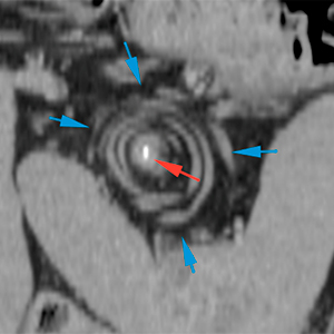
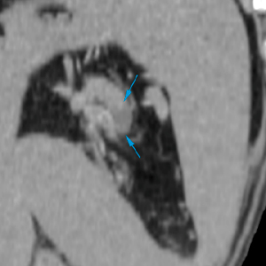
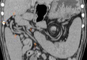
At level of the splenic pedicle, caudal to the stomach, there is a whirl sign (blue arrows), with a hyperattenuating area (298 UH) in its central portion (red arrow), consistent with a splenic torsion.
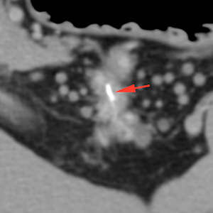
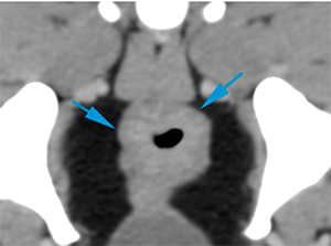
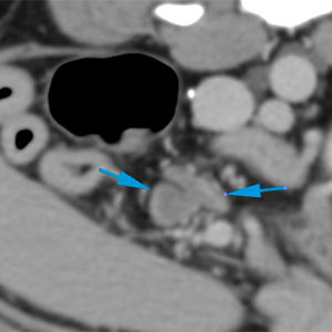
In the post-contrast study, the splenic parenchyma, as well as part of the splenic vasculature, shows a mild/moderate contrast enhancement (pre-contrast 60 UH; post-contrast 80 UH). In the area of the splenic hilum, there is a filling defect within the vessels, which could be consistent with a region of thrombosis (blue arrow). Additionally, there are multiple vessels with a tortuous path, distributed throughout the cranial aspect of the peritoneum (orange arrows).
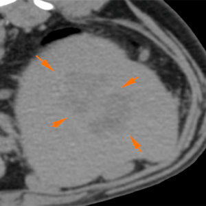
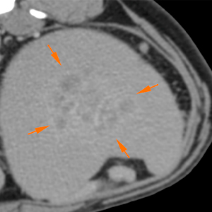
In the left caudal aspect of the spleen, there is a heterogeneous lesion, with ill-defined margins, soft tissue attenuation and hypoattenuating compared to the rest of the splenic parenchyma (orange arrows). In the post-contrast study, the lesion shows a moderated contrast enhancement, mainly peripheral, remaining hypoattenuating compared to the rest of the spleen.
Pre-contrast
Post-contrast
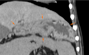
The stomach has a normal position, although displaced cranially due to the mass effect generated by the spleen (orange arrows). The intestinal loops are also caudally displaced (pink arrows). There is a diffuse and moderate thickening of the colonic wall, being more evident in its descending portion (blue arrows).
The jejunal lymph nodes are moderately enlarged, with a slightly heterogenous contrast enhancement (blue arrows).
Diagnosis
Findings consistent with splenic torsion.
Possible thrombosis along the splenic pedicle, as well as presence of collateral vascularization vs congestion of all the adjacent vasculature.
Lesion at the left caudal aspect of the spleen, most likely consistent with a malignant lesion (neoplastic – histiocytic sarcoma, among others, given the results of the cytology), although a benign lesion (ischaemia, necrosis …) is also possible.
Moderate diffuse thickening of the wall of the colon, consistent with inflammatory changes (colitis), most likely. Other differentials, such as diffuse neoplasia (lymphoma), are less likely.
Moderate jejunal lymphadenopathy, most likely reactive.
Comments
Splenectomy and histopathological analysis of the spleen are recommended in order to reach a definitive diagnosis.
(Mon. to Fri. 9 a.m. to 6 p.m. gmt+1) Welcome, How can we help you?
We use cookies to ensure that we give you the best experience on our website. If you continue to use this site we will assume that you are happy with it.Ok


No comment yet, add your voice below!