8-years-old female boxer with pelvic limbs weakness and leishmaniosis. In the radiographic study of the lumbar spine, a soft tissue lesion with mineralizations at the ventral aspect of the vertebral bodies of L7-S1 was seen.
An abdominal CT was performed.
Description
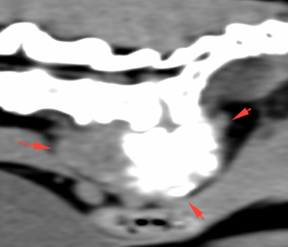
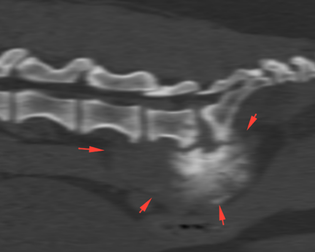
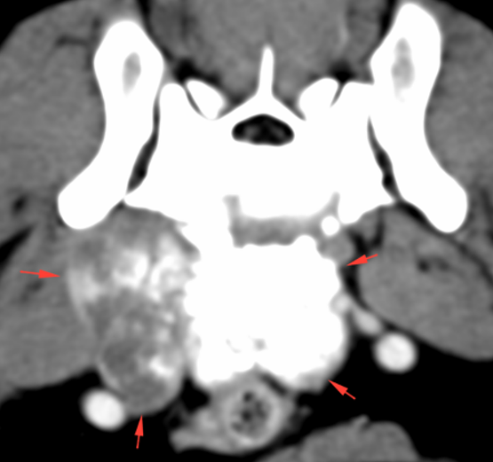
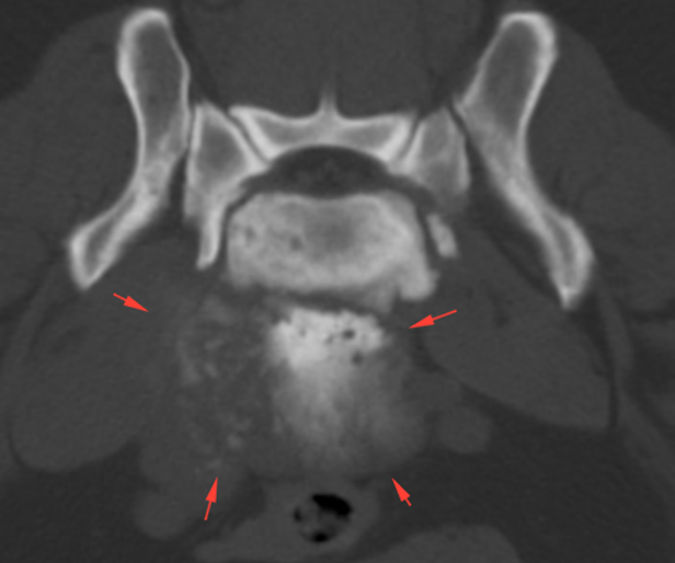
There is an ill-defined and heterogeneous soft tissue mass in the ventral aspect of the lumbosacral region (red arrows). This lesion seems to originate from the ventral aspect of the vertebral bodies of L7-S1 and hypaxial musculature, affecting the psoas major and minor muscles. The mass has an amorphous bone attenuation component. In the post-contrast series, the soft tissue component of the mass shows a marked heterogeneous contrast enhancement. The osseous structures do not show evident abnormalities at this level.
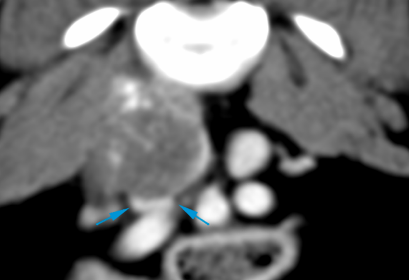
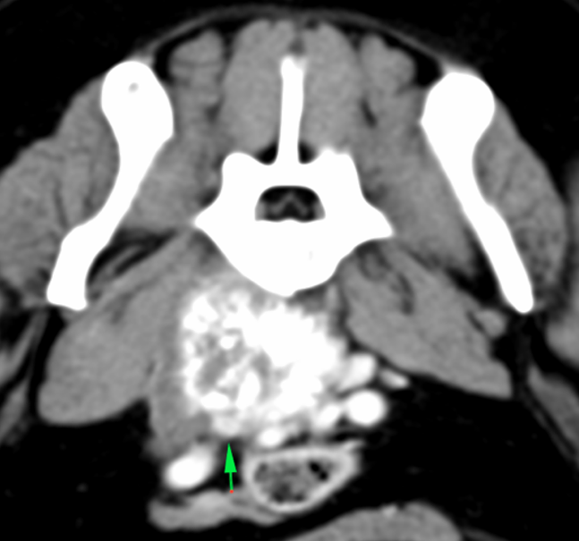
The mass causes a moderate mass effect displacing ventrally the major abdominal vessels (caudal vena cava and aorta), with marked compression of the right common iliac vein (blue arrows). The most caudal portion of the latest is embedded within the mass (green arrow), without being clearly visible at this level. More caudally, the external iliac vein can be seen, with normal appearance.
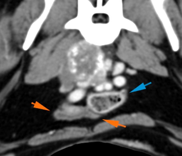
There is ventral displacement of the urinary bladder’s neck (orange arrows) and colon (blue arrows).
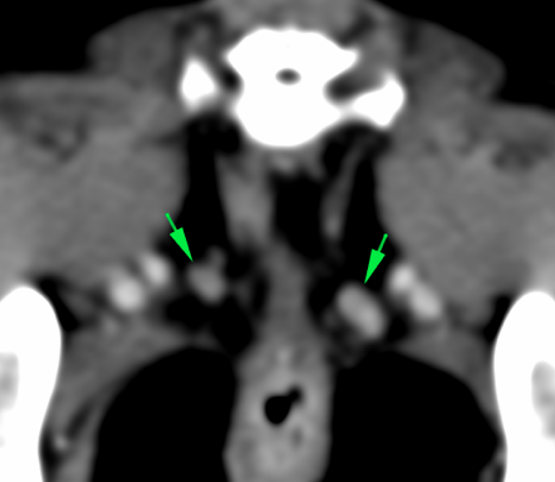
The sacral lymph nodes are mildly enlarged (left > right) with homogeneous contrast enhancement (green arrows). The left medial and internal iliac lymph nodes are normal. However, the right lymph nodes are not clearly visualized because of the mass.
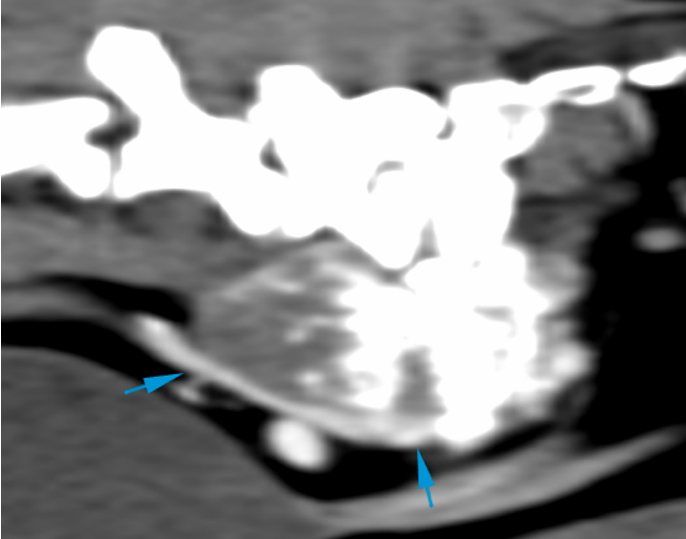
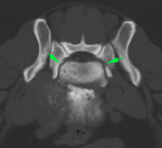
There is spondylosis deformans in the lumbosacral region extending laterally and dorsally with narrowing of the L7-S1 intervertebral foraminae (green arrows).
Diagnosis
Sublumbar mass, with mixed bone and soft tissue components, related to the vertebral bodies of L7-S1, affecting the hypaxial musculature. This is most likely consistent with neoplasia (sarcoma – sublumbar extra-skeletal osteosarcoma or other sarcomas). Benign bone lesions or granulomatous lesions are unlikely given its characteristics.
Compression of the common iliac vein due to the mass. Invasion of this vessel by the mass cannot be ruled out.
Sacral lymphadenopathy (left > right): metastatic vs reactive
Mild spondylosis deformans and stenosis of both L7-S1 intervertebral foraminae – degenerative and incidental finding most likely, but nerve root compression cannot be ruled out -.
Comments
Fine needle aspiration of the mass is recommended in order to reach a definitive diagnosis. Ultrasound evaluation of the vasculature adjacent to the mass could be considered. The mass and the affection of the vasculature or the changes at the lumbosacral level could explain the patient’s clinical signs; nerve involvement at this level cannot be ruled out.
(Mon. to Fri. 9 a.m. to 6 p.m. gmt+1) Welcome, How can we help you?
We use cookies to ensure that we give you the best experience on our website. If you continue to use this site we will assume that you are happy with it.Ok


No comment yet, add your voice below!