5-year-old female Yorkshire Terrier. She already came previously for an abscess that was oozing in the right frontal area. Treatment was administered but since the abscess did not resolve, lavages were done and a drain was placed. Ultrasound was performed and an encapsulated abscess was observed without structures consistent with foreign bodies.
Region: Skull, pre and post contrast.
Description
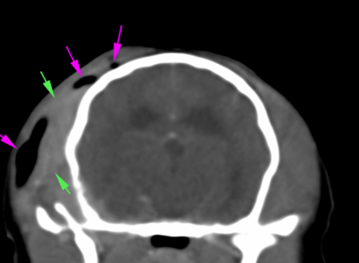
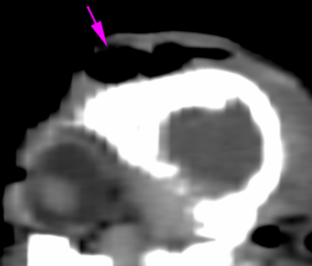
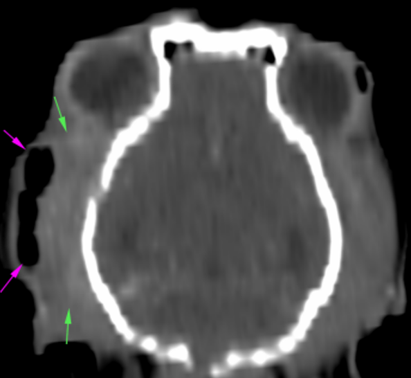
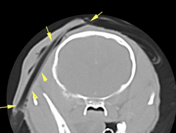
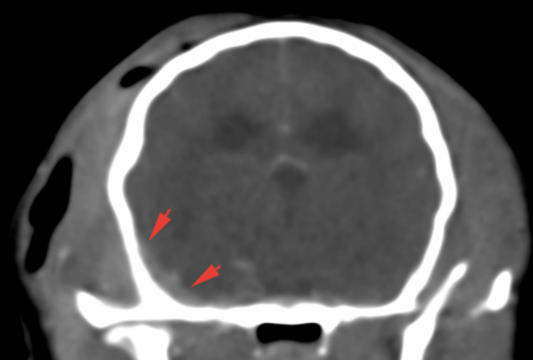
There is an extensive lesion with ill-defined margins, which originates immediately caudal to the right orbit and extends caudally through the parietal and temporal region, affecting the masticatory musculature at this level (temporalis muscle). There is thickening/bulging of the soft tissues, with presence of gas inside (subcutaneous emphysema) (pink arrows) and, the right temporalis muscle shows a marked and slightly heterogeneous contrast enhancement (green arrows). No structures consistent with foreign bodies can be seen observed in the lesion. At this level, there is a drainage catheter (described in the history) (yellow arrows). Both ocular globes and retrobulbar spaces are intact.
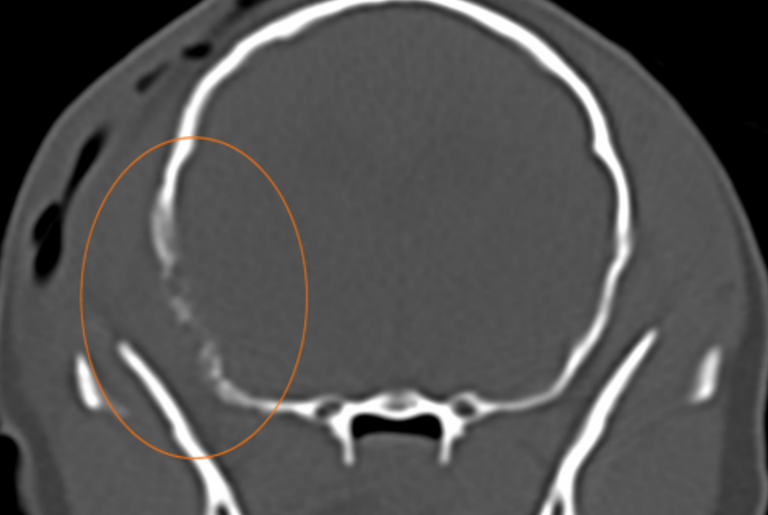
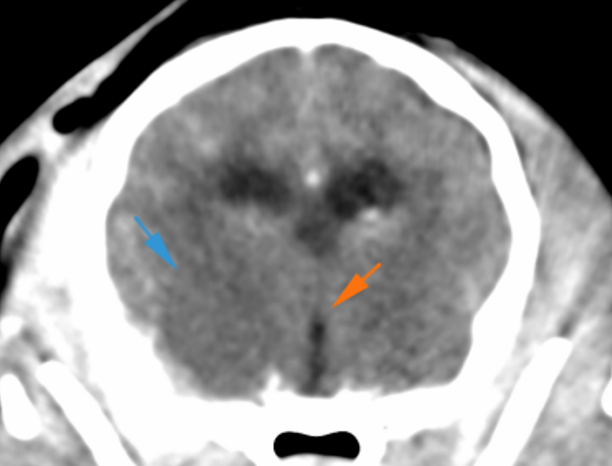
Adjacent to the above-described lesion, and caudal to the right temporomandibular joint, a moderate permeative and moth-eaten type osteolysis is visible, affecting the temporal bone (orange circle), which shows an irregular surface. The temporomandibular joint is intact. Associated with this level and intracranially, there is marked thickening and enhancement of the meninges (red arrows); as well as a hypoattenuating intra-axial lesion, with ill-defined margins, in the region of the right piriform lobe, affecting mainly the white matter (blue arrow), which does not show post-contrast enhancement, most likely consistent with vasogenic oedema. This lesion shows a mild associated mass effect, minimally displacing the ventral portion of the 3rd ventricle towards the left (orange arrows). However, there are no signs of transtentorial or cerebellar herniation.
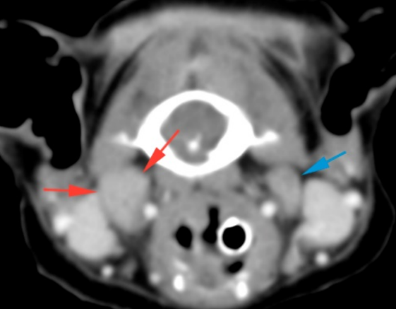
The right medial retropharyngeal lymph node (red arrows) is enlarged and asymmetrical compared to the contralateral side (blue arrow).
Diagnosis
Lesion at the right frontoparietal level (temporal muscle) consistent with an inflammatory/infectious lesion (abscess) most likely secondary to penetrating injury/foreign body (although not visible on the CT scan). Associated subcutaneous emphysema and marked myositis of the right temporalis muscle.
Aggressive osseous lesion of the adjacent temporal bone, which is consistent with associated osteomyelitis. Other differentials, such as a neoplastic process, are unlikely.
Intracranial extension of this lesion with thickening of the meninges and an area of vasogenic oedema in the right temporal area, consistent with meningoencephalitis, most likely, with a mild mass-effect towards the left.
Moderate right medial retropharyngeal lymphadenopathy, most likely reactive.
Comments
The lesion described at the level of the right temporal muscle is consistent with an infectious lesion (abscess and associated myositis), which is probably secondary to a foreign body, even if not visible on the CT study, or secondary to another penetrating injury. Consider that intracranial extension of this process is visible with possible osteomyelitis of the temporal bone and associated meningoencephalitis. Sampling of the temporal lesion to perform a culture is advised in order to achieve a definitive diagnosis.
(Mon. to Fri. 9 a.m. to 6 p.m. gmt+1) Welcome, How can we help you?
We use cookies to ensure that we give you the best experience on our website. If you continue to use this site we will assume that you are happy with it.Ok


No comment yet, add your voice below!