Thoracic spinal mass with extradural extension in a dog
5-year-old male German Shepperd dog with pelvic limbs weakness. A mass at the level of thoracic vertebrae is detected during the general physical examination.
A thoracic spine CT is performed.
A thoracolumbar spine and thoracic CT scan was performed.
Description
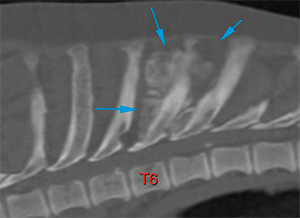
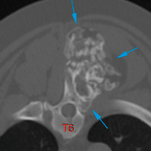
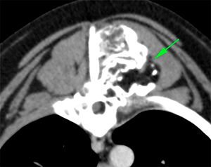
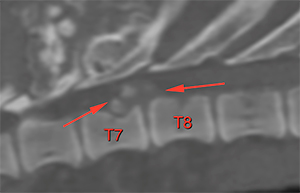
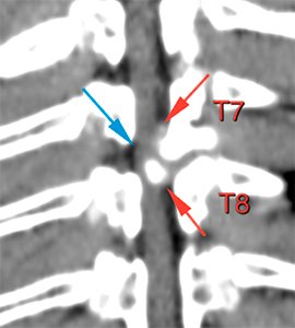
There is a mineral attenuating mass, with solid and amorphous new bone formation affecting the spinous processes of T6 and T7 (blue arrows) occupying the left epaxial region. This lesion shows multiple fat attenuating areas (green arrow).
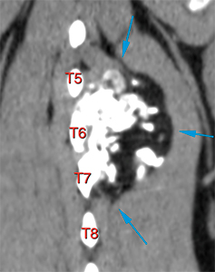
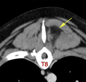
This mass extends from the caudal aspect of the spinous process of T5 to the caudal aspect of the vertebral body of T8 (blue arrows). No abnormalities are seen at the spinous process of T8, but part of the fat attenuating component of the mass is adjacent to it, infiltrating the left epaxial musculature (yellow arrow).
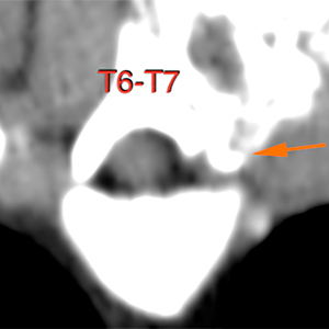
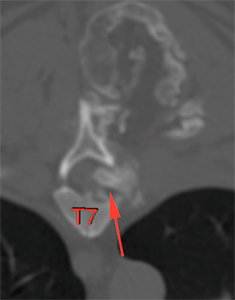
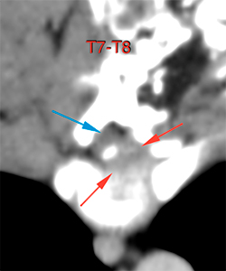
The mass protrudes into the region of the left intervertebral foramen, at the level of T6-T7 (orange arrow), and enters the left intervertebral foramen of T7-T8 as an extradural lesion, occupying the vertebral canal. It extends caudally along the ventral and the left lateral aspect of the canal up to the cranial aspect of T8 (red arrows). This lesion causes a marked compression and displacement of the spinal cord (blue arrow), occupying up to 70% of the diameter of the vertebral canal at the level of the intervertebral space of T7-T8.
Diagnosis
Thoracic spinal mass affecting the spinous processes and the dorsal lamina of the vertebral bodies of T6 and T7, with bone, soft tissue and fat attenuating components. This is most likely consistent with a neoplastic process like chondrosarcoma; other neoplasia, such as osteosarcoma, cannot be ruled out. A benign neoplastic process, such as osteochondroma, cannot be excluded; a malignant transformation of said process into chondrosarcoma is also possible.
Left extradural extension of the mass causing a moderate/severe compressive myelopathy in T7-T8.
Comments
Biopsy of the mass with histopathological analysis is recommended in order to reach a definitive diagnosis. Consider that the patient’s clinical signs are probably related to compressive myelopathy secondary to extradural extension of the lesion.
(Mon. to Fri. 9 a.m. to 6 p.m. gmt+1) Welcome, How can we help you?
We use cookies to ensure that we give you the best experience on our website. If you continue to use this site we will assume that you are happy with it.Ok


No comment yet, add your voice below!