17-year-old female domestic short hair. Chronic cough since she was 8 years old. Currently, inspiratory and expiratory dyspnoea.
A thoracic CT scan was performed.
Description
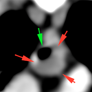
There is a broad-base well-defined soft tissue mass (1,6 cm CrCd x 0,5 cm DV x 0,8 cm LM) at the ventral aspect of the caudal tracheal lumen (red arrows). This lesion shows a mild diffuse contrast enhancement in the post-contrast series. At its cranial portion, the mass seems to obliterate the tracheal lumen, narrowing its lumen by 80% (green arrow).
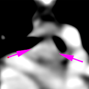
This lesion reaches the carina and there is narrowing of the ventral aspect of both mainstem bronchi (pink arrows).
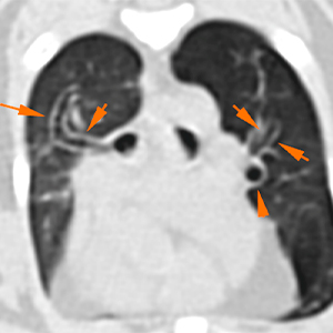
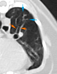
The bronchial walls are mildly/moderately thickened, with cylindrical bronchiectasis (orange arrows). Mild tree-in-bud lesions near the bronchi are seen (blue arrows).
Diagnosis
Intratracheal mass with narrowing of the caudal thoracic tracheal and mainstem bronchi lumen, most likely consistent with neoplasia (lymphoma, carcinoma…). Other differentials, such as polyp or granuloma, are less likely.
Thickening of the bronchial walls with bronchiectasis and tree-in-bud pattern: feline asthma, most likely.
Comments
Bronchoscopy and sampling of the tracheal mass +/- bronchoalveolar lavage are recommended.
(Mon. to Fri. 9 a.m. to 6 p.m. gmt+1) Welcome, How can we help you?
We use cookies to ensure that we give you the best experience on our website. If you continue to use this site we will assume that you are happy with it.Ok


No comment yet, add your voice below!