Transposition of caudal vena cava with retrocaval ureter in a dog
5-year-old male Greyhound with no symptoms. In the abdominal ultrasound, prior to his castration, hydronephrosis and hydroureter were observed.
An abdominal CT scan was performed.
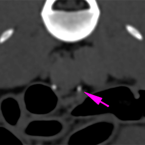
A thoracolumbar spine and thoracic CT scan was performed.
Description
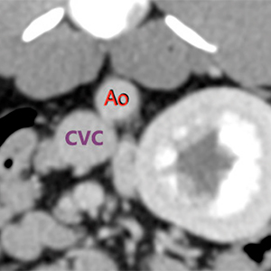
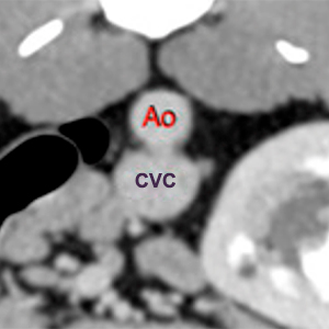
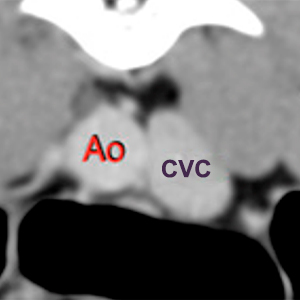
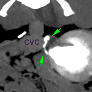
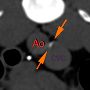
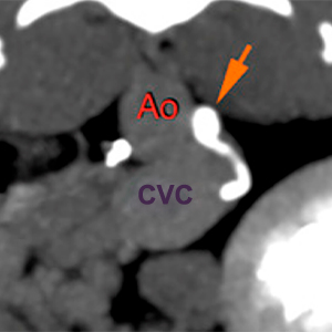
There is a transposition of the caudal aspect of the caudal vena cava (CVC), affecting its pre-renal portion, which shows a marked ventrolateral deviation, being located in the left lateral aspect of the aorta (AO) transition area.
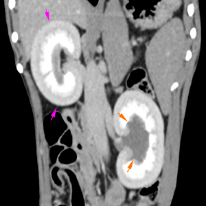
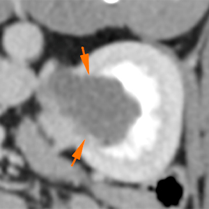
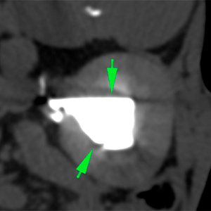
The left renal pelvis is moderately/markedly distended, filled with hypoattenuating content (orange arrows) and, during the excretory urography, homogeneously filled with contrast (green arrows). Nevertheless, the left kidney has a normal size and shape, with good corticomedullar differentiation. The right kidney has a normal size and shape, without signs of distention of the pelvis (pink arrows).
The proximal portion of the left ureter is moderately distended, with a slightly tortuous path, surrounding the CVC (green arrows), located dorsal to the latest and passing between the aorta and the CVC, where it is compressed (orange arrows). More caudally, the left ureter presents a normal position and diameter (located ventrally to the large vessels) (pink arrows), inserting into a normal ureterovesical union. The left ureter has a normal diameter and path, inserting at level of the trigone of the urinary bladder.
Diagnosis
Moderate hydronephrosis with mild left hydroureter, secondary to transposition of the caudal vena and retrocaval ureter, with compression of the latest.
In spite of the hydronephrosis, the left kidney shows a normal morphology and architecture.
Comments
The findings are consistent with transposition of the caudal aspect of the CVC due to a congenital vascular anomaly, with presence of left retrocaval ureter, compressed between the aorta and CVC, causing a mild hydroureter and a moderate left hydronephrosis.
(Mon. to Fri. 9 a.m. to 6 p.m. gmt+1) Welcome, How can we help you?
We use cookies to ensure that we give you the best experience on our website. If you continue to use this site we will assume that you are happy with it.Ok

No comment yet, add your voice below!